
Case presentation
A 21-year-old female of Italian origin presented to the emergency department with a one-week history of a swollen, increasingly painful left leg. The swelling involved the whole leg from groin downwards and was associated with heat and erythema. She had no pleuritic chest pain or breathlessness, and was otherwise systemically well. There was no past history of leg swelling, and no history of recent travel, surgery or trauma nor significant family history. She had recently stopped the combined oral contraceptive pill one-month prior to admission. On examination she was alert but in discomfort, apyrexial and haemodynamically stable. Cardiovascular, respiratory and abdominal examination was unremarkable. Body mass index (BMI) was 40 kg/m2. Her right leg was normal. Her left leg was markedly swollen, erythematous, hot and tender to palpation from the groin downwards. Pulses were difficult to feel. There was no sensory loss and no muscular weakness. Calf veins were tender. Initial investigations showed a normal full blood count, renal function and clotting. Chest radiograph showed clear lung fields, normal heart and mediastinum. Urinalysis was negative for nitrates, leucocytes, blood and protein.
What is the differential diagnosis and likely diagnosis?
The most likely diagnosis in a swollen tender leg is that of a deep vein thrombosis (DVT). Leg swelling is, however, a common presentation and can have systemic or local aetiologies. Systemic causes such as cardiac or renal failure are more likely to cause bilateral limb swelling, would be unlikely in a patient of this age and are likely to be associated with other examination findings or abnormalities on chest radiography.
Lymphoedema is an infrequent cause of leg swelling. Our patient had no history of previous surgery and the acute nature of onset makes the diagnosis unlikely. A further differential would be infective and inflammatory causes such as cellulitis, myositis or fasciitis. Systemic features of infection would be expected (pyrexia, raised white cell count), however, and no obvious portal of infection was noted.
DVT was the primary differential but with uncertain aetiology. The thrombus appeared clinically to be very proximal and extensive and the patient had no clear risk factors for venous thromboembolism apart from previous use of the oral contraceptive pill and being obese. A possible predisposition to thrombosis, such as thrombophilia, or a pelvic mass were considered. As there was severe swelling and pain, and pulses were difficult to palpate, a compartment syndrome was considered.
What was the initial management?
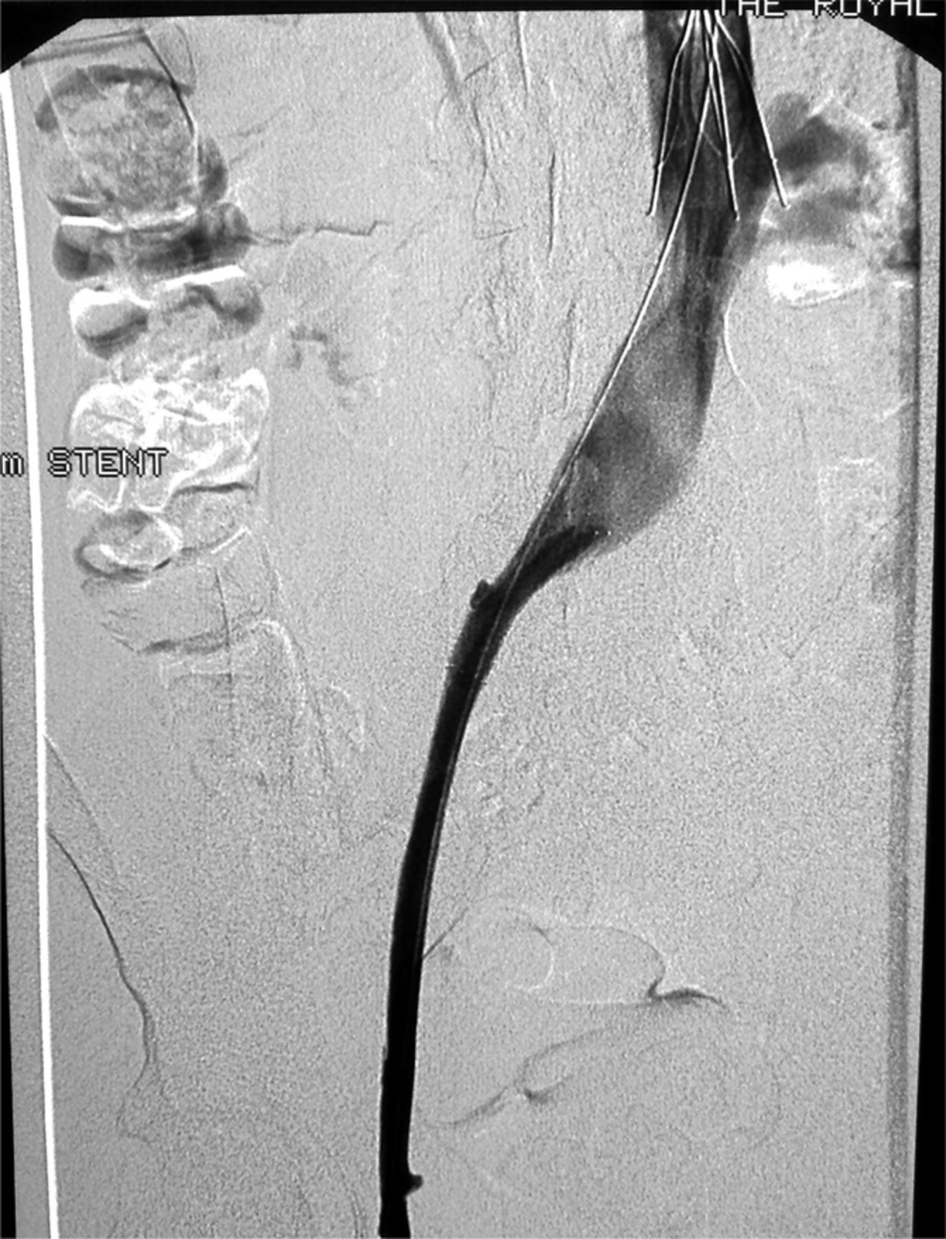
Due to the high clinical suspicion of DVT immediate anti-coagulation was commenced with low molecular weight heparin in accordance with current guidelines, and analgesia was prescribed.1 Doppler ultrasonography of the leg and pelvis revealed an extensive left femoral vein DVT, extending into the pelvis and possibly into the iliac vein, although it was impossible to determine the proximal extent of the thrombus due to the patient's BMI. Computed tomography (CT) of the abdomen and pelvis showed gross swelling and oedema within the left thigh and the veins were expanded with clot. The thrombus extended up to the point of origin of the left common iliac vein (Fig 1). The right common iliac artery crosses anterior to the vein in this region (May – Thurner syndrome).2 There was no inferior vena cava (IVC) thrombus demonstrated. Thrombophilia screen was negative.

Image demonstrating extensive clot within the iliac vein. Inferior vena cava filter in place.
Case progression
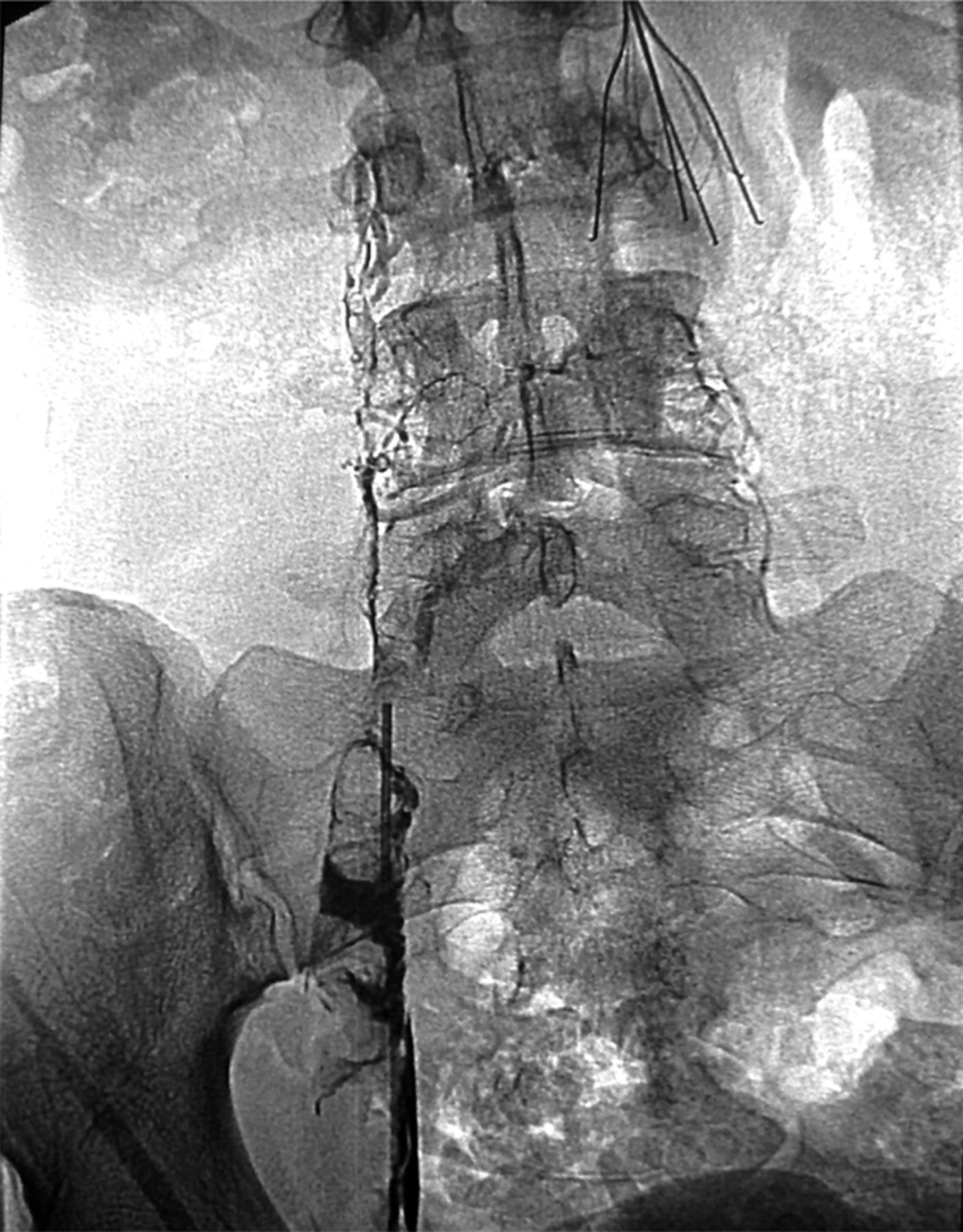
Due to the extensive size of the thrombus, and high risk of post-thrombotic syndrome leading to chronic venous sequelae, her case was discussed immediately with the haematologists and interventional radiologists. The risks and benefits of pharmaco-mechanical localised thrombolysis (catheter-directed thrombolysis therapy) were discussed carefully with the patient, and she agreed to undergo the procedure. An IVC filter was placed through the right internal jugular vein positioned at L2/3 to prevent pulmonary emboli during the procedure. Subsequently, access was gained to the popliteal vein, catheter-directed pharmacological thrombolysis with recombinant tissue plasminogen activator (tPA) was administered, and subsequent mechanical disruption of the thrombus was undertaken. The stenosis of the left common iliac vein was stented, and the patient received a continuous infusion of tPA for two hours post-procedure, resulting in a widely patent femoral and iliac vein (Fig 2). She was observed overnight on the high dependency unit. The patient's pain and swelling considerably improved within 24 hours. The IVC filter was removed seven days post-procedure, and the patient anti-coagulated with warfarin, with a plan for six months of anticoagulation and haematological follow-up.
Post-catheter-directed pharmacological and mechanical thrombolysis. The vein is now patent with iliac vein stent in place.
Discussion
May – Thurner syndrome is an uncommon clinical scenario, describing the compression of the left common iliac vein by the right common iliac artery. The resulting stenosis leads to venous stasis, and may progress to leg swelling, varicosities, DVT, pulmonary embolism or phlegmasia cerulea dolens. The extensive DVT seen in this condition can have serious sequelae including post-thrombotic syndrome and pulmonary embolism, although the latter is less common due to the stenosis of the vein. It is therefore important not to delay treatment.
The prevalence of May – Thurner syndrome is unknown, but estimated to occur in 2–5% of patients undergoing investigation for lower extremity venous disorders.3 The condition is more commonly seen in overweight females, typically in their late teens and early 20s. Partial obstruction of the left iliac vein occurs due to the physical entrapment from the right iliac artery and intimal hypertrophy of the vein from the pulsatile force of the right iliac artery. Diagnosis is made either by iliac venography to demonstrate both the compression and pressure gradient or contrast enhanced CT.
The treatment of May – Thurner usually involves interventional methods to relieve the obstruction in addition to anticoagulation. Surgical thrombectomy followed by iliocaval stenting has been used previously.4 More recently, catheter-directed thrombolytic therapy, angioplasty, stenting together with IVC filters have been used successfully.5,6 The use of IVC filters is controversial, as the risk of pulmonary embolus is small, when thrombolytic therapy is used.
While DVT is a very common clinical scenario, this case highlights the fact that extensive unexplained DVT requires further investigation and treatment with thrombolysis should be considered, particularly in younger patients, as it may have significant benefits in terms of reduced risk of complications in the future.
Key learning points

- © 2010 Royal College of Physicians


Jump to section
Related Articles
Cited By...
- No citing articles found.