
Key learning points
RS3PE presents mainly in men over 50 years old with bilateral hand and foot synovitis and pitting oedema.
Investigations usually show normal radiography and negative rheumatoid factor.
There are possible associations with malignancy and other rheumatological conditions, especially polymyalgia rehumetica.
The condition responds well to low-dose steroids.
Case presentation
A 78-year-old man with osteoarthritis, hypertension, and chronic obstructive pulmonary disease (COPD) initially presented to the emergency department (ED) with right, and to a lesser extent, left hand swelling, of five weeks' duration. He also described stiffness in his hands, especially in the morning, as well as bilateral shoulder pain. He was given a two-week course of antibiotics (clindamycin and erythromycin) by his GP for a presumed cellulitis, and also tried a one-week course of prednisolone 20 mg. There had been no resolution in symptoms.
On presentation to the ED, he was noted to have a diffusely swollen right, and mildly swollen left hand. All pulses were present. No other clinical abnormalities were found. Bloods showed a normal biochemical profile and urate, but C-reactive protein (CRP) was mildly elevated at 36 mg/l with erythrocyte sedimentation rate (ESR) 20 mm/hr. Radiography of the hands showed no erosions or calcification and an ultrasound doppler of his right arm ruled out thrombosis. He was discharged for review to the rheumatology outpatients department, pending autoantibodies and magnetic resonance imaging (MRI) of his hands.
One week later, the patient re-presented with further swelling and pain in his hands. Examination on this occasion showed pitting oedema on the dorsal aspect of the right hand. Movement of the fingers was limited by the swelling but sensation was normal. Pulses were present with a capillary refill of two seconds. Blood results are shown in Table 1.
Blood results on the second admission.
What is the differential diagnosis and the most likely diagnosis?
On initial presentation, the patient had already had a two-week course of antibiotics in the community. A resolving cellulitis of the right hand may fit with the elevated CRP, although the left hand was also slightly swollen. Late onset rheumatoid arthritis should be considered, although the negative rheumatoid factor and normal radiograph would be unusual. Gout remains a possibility despite the normal urate level. The bilateral shoulder pain may suggest polymyalgia rheumatica (PMR), but this does not explain the hand swelling. Some improvement with the prednisolone would have been expected with the above diagnoses, although only a short course was trialled.
At representation, pitting oedema of the right hand was noted and the inflammatory markers were raised. Septic arthritis should be considered, or possibly a relapsing cellulitis of the right hand or secondary skin infection complicating a primary pathology. The pitting oedema makes lymphoedema a possibility. Scleroderma can also present with swelling and oedema may explain the swelling of both hands. Remitting seronegative symmetrical synovitis with pitting oedema (RS3PE), a little known diagnosis, may fit with the overall presentation.
What is the initial management?
Exclusion of upper limb thrombus is required, and treatment with empirical antibiotics may be considered, if infection is thought to be a realistic possibility. Elevation of the swollen limb should be undertaken. Autoantibodies should be sent to guide any rheumatological diagnosis and MRI of the hand, to look for soft tissue infection as well as other structural abnormalities, performed.
Case progression
The hand swelling reduced somewhat with intravenous antibiotics, but the MRI showed no evidence of cellulitis. The patient was discharged on oral antibiotics, with rheumatological follow-up. Two days later, the patient was found collapsed at home. On examination, he was short of breath and appeared cyanotic. Pulse rate was 110 bpm and regular, blood pressure 135/75 mmHg. Further cardiorespiratory examination was normal. His hands were noted to be more swollen, with synovitis of the small joints and ongoing pitting oedema.
A computed tomography (CT) pulmonary angiogram showed a pulmonary embolus (PE) and he was anti-coagulated. Abdominal CT showed a cystic mass in the pancreas (Fig 1). Fluid from the mass was aspirated and proved non-diagnostic. Conservative management with serial imaging was advised by the hepatobiliary surgeons.
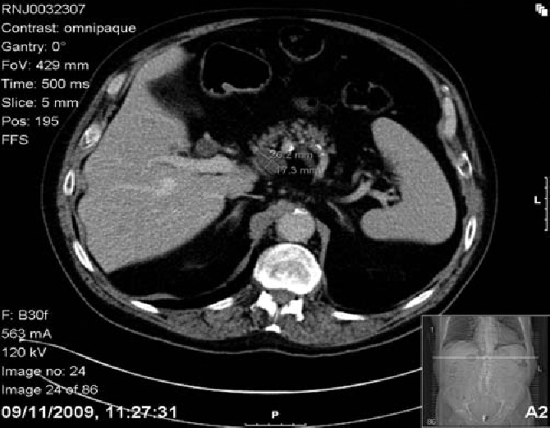
Computed tomography scan of the abdomen showing a cystic mass in the pancreas.
A clinical diagnosis of RS3PE was made by the rheumatologists and the patient was started on prednisolone 20 mg daily. The swelling reduced considerably over the next few weeks and the steroid dose weaned.
Discussion
First described by McCarty et al. in 1985,1 RS3PE is a condition which presents with acutely swollen hands and often feet. Classically, there is synovitis and tenosynovitis with pitting oedema of the dorsum of the hand. It occurs mainly in men over the age of 50 years old. Radiographs are usually normal and rheumatoid factor is negative but inflammatory markers may be elevated. Resolution of symptoms occurs with low-dose steroids.
A number of cases of RS3PE are associated with malignancy.2 There are also reports of RS3PE in association with most other rheumatological conditions, including rheumatoid arthritis, spondyloarthropathies, psoriatic arthritis and PMR.3 Cases of PMR often have distal signs and more frequently distal relapses. Therefore, RS3PE has been hypothesised to be part of a spectrum of PMR, although RS3PE does not require such high doses of steroids.4
In the case above, the patient initially presented with hand swelling. He also described shoulder girdle pain, which hints at a diagnosis of PMR. It is likely that the 20-mg course of prednisolone given by the general practitioner, if continued for a longer duration, would have considerably improved the hand swelling. A higher dose of steroids would have been needed to control the symptoms of PMR. It could also be speculated that the pancreatic mass and RS3PE are not coincidental, although as yet the mass has not been confirmed as malignant.
RS3PE is a rare diagnosis and remains only partially understood. The case presented above would not be an unusual course of events for someone presenting with RS3PE. Acute physicians should be aware of the existence of this rheumatological condition, as well as its sometimes serious associations.
- Royal College of Physicians


Jump to section
Related Articles
Cited By...
- No citing articles found.