
Main menu

  • Home
  • Our journals
    • Clinical Medicine
    • Future Healthcare Journal
  • Subject collections
  • About the RCP
  • Contact us

Clinical Medicine Journal

  • ClinMed Home
  • Content
    • Current
    • Ahead of print
    • Archive
  • Author guidance
    • Instructions for authors
    • Submit online
  • About ClinMed
    • Scope
    • Editorial board
    • Policies
    • Information for reviewers
    • Advertising

User menu

  • Log in

Search

  • Advanced search
RCP Journals
Home
  • Log in
  • Home
  • Our journals
    • Clinical Medicine
    • Future Healthcare Journal
  • Subject collections
  • About the RCP
  • Contact us
Advanced

Clinical Medicine Journal

clinmedicine Logo
  • ClinMed Home
  • Content
    • Current
    • Ahead of print
    • Archive
  • Author guidance
    • Instructions for authors
    • Submit online
  • About ClinMed
    • Scope
    • Editorial board
    • Policies
    • Information for reviewers
    • Advertising

Homonymous left hemianopia secondary to traumatic extracranial vertebral artery dissection

Hala Kanona, Vanessa Tuboku-Metzger and Khaldoon Wahab
Download PDF
DOI: https://doi.org/10.7861/clinmedicine.11-6-623
Clin Med December 2011
Hala Kanona
Heart of England Foundation Trust, Good Hope Hospital, Sutton Coldfield, Birmingham
Roles: Core surgical trainee
  • Find this author on Google Scholar
  • Find this author on PubMed
  • Search for this author on this site
  • For correspondence: hkanona@yahoo.co.uk
Vanessa Tuboku-Metzger
Heart of England Foundation Trust, Good Hope Hospital, Sutton Coldfield, Birmingham
Roles: Core surgical trainee
  • Find this author on Google Scholar
  • Find this author on PubMed
  • Search for this author on this site
Khaldoon Wahab
Heart of England Foundation Trust, Good Hope Hospital, Sutton Coldfield, Birmingham
Roles: Consultant orthopaedic surgeon
  • Find this author on Google Scholar
  • Find this author on PubMed
  • Search for this author on this site
  • Article
  • Figures & Data
  • Info & Metrics
Loading

Abstract

This lesson reports the case of a 32-year-old female presenting with left-sided homonymous hemianopia after a road traffic accident. A diagnosis of vertebral artery dissection (VAD) was missed at presentation after an initial computed tomography head scan was normal. Later, VAD was confirmed on neck magnetic resonance imaging. VAD is an uncommon cause of stroke in the young and, to date, there are no current emergency guidelines available for the management of patients with the condition.

Introduction

Traumatic vertebral artery dissection (VAD) is frequently unrecognised.1 It accounts for 20% of strokes in patients aged 45 and younger2 and up to 8% of those aged 60 and younger.3 It occurs in about 1% of patients following a blunt mechanism of injury.2 Dissection of the extracranial vertebral or carotid arteries are a relatively common cause of ischaemic stroke in the young.3,4 Dissection can cause ischaemic or haemorrhagic stroke although the latter is much more uncommon.5

The common symptoms of VAD at presentation include headache, vertigo, paraesthesia, neck and occipital pain.3 In addition, there is evidence from the existing literature describing ophthalmic symptoms occurring as the primary presenting complaint in 15–86% of cases.6,7 This lesson describes the case of a 32-year-old female who presented with left homonymous hemianopia secondary to traumatic VAD.

Lesson

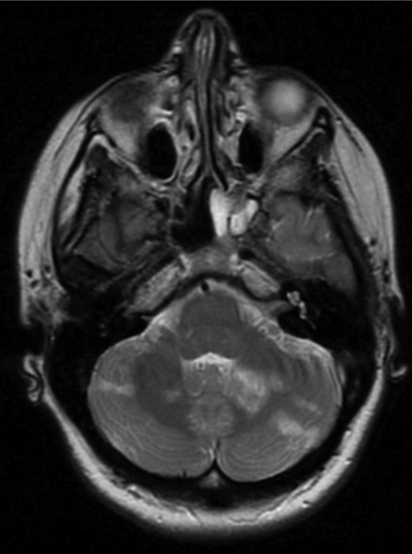
Following a road traffic accident, a 32-year-old patient presented to accident and emergency with frontal headache radiating to the occiput, blurred vision, memory loss and neck pain. Initial examination revealed the patient to be haemodynamically normal. There was no spinal or abdominal tenderness and neurovascular examination was initially normal. Upon further examination she was found to have a left homonymous hemianopia. Magnetic resonance imaging (MRI) of the head showed evidence of multiple foci of signal abnormality in the cerebellum (Fig 1) and right temporal lobe with evidence of infarction in the right occipital lobe and smaller areas of signal abnormality in the left occipital lobe (Fig 2). Normal flow-voids were seen within the vertebral and basilar arteries with marginal enhancement. No evidence of any carotid abnormality was found on ultrasound scanning. Subsequent MRI imaging of the neck confirmed the suspected diagnosis of vertebral artery dissection, showing a narrowed segment of the right vertebral artery around C7. The patient was commenced on aspirin and dipyramidole and was discharged with follow-up under the neurologists. Ten months after the initial injury, the patient still has persisting visual impairment.

Fig 1.
  • Download figure
  • Open in new tab
  • Download powerpoint
Fig 1.

T2-weighted magnetic resonance image of the brain–multiple foci of abnormal enhancements are visible throughout both cerebellar hemispheres.

Fig 2.
  • Download figure
  • Open in new tab
  • Download powerpoint
Fig 2.

T2-weighted magnetic resonance image of the brain–showing foci of abnormal enhancement in the right occipital lobe.

Discussion

While VAD can occur spontaneously, some studies have shown that it occurs in relation to mild or significant trauma in up to 83% of cases.3 A literature review conducted between 1990 and 2004 showed that the main mechanisms of injury associated with VAD included: distraction/extension, distraction/flexion and lateral flexion injuries.8 VAD has been reported as a rare consequence of whiplash9 as illustrated in this case report. Due to the wide and variable symptoms, VAD is commonly misdiagnosed as musculoskeletal pain10 at presentation which can lead to a delay in treatment. Commonly vertebral artery dissection is diagnosed using MRI T1-suppressed axial images.11 It is generally accepted that VAD leading to ischaemic stroke is managed by anticoagulation10 or antiplatelet therapy.

Conclusion

VAD is a relatively common cause of stroke in the young and can present with a wide variety of musculoskeletal and neurological symptoms. Awareness of the condition is important in assuring that the diagnosis is not missed. Thorough neurological examination (including visual field testing) with appropriate imaging is essential in diagnosing those patients with suspected VAD. Neglecting these steps can delay treatment of patients and affect prognosis. To date, there are no current emergency guidelines available for the management of patients with VAD.

  • © 2011 Royal College of Physicians

References

  1. ↵
    1. Ribbons T,
    2. Bell S
    . Neck pain and minor trauma: normal radiographs do not always exclude serious pathology. Emerg Med J 2008; 25: 609–10doi:10.1136/emj.2007.050328
    OpenUrlAbstract/FREE Full Text
  2. ↵
    1. Redekop GJ
    . Extracranial carotid and vertebral artery dissection: a review. Can J Neurol Sci 2008; 35: 146–52
    OpenUrlPubMed
  3. ↵
    1. Bartels E
    . Dissection of the extracranial vertebral artery: clinical findings and early noninvasive diagnosis in 24 patients. J Neuroimaging 2005; 16: 24–33doi:10.1177/1051228405280646
    OpenUrlCrossRef
  4. ↵
    1. Betram M,
    2. Ringleb P,
    3. Fiebach J,
    4. et al
    . Spectrum of neurological symptoms in dissections of brain-supplying arteries. Dtsch Med Wochenschr 1999; 124: 273–8
    OpenUrlPubMed
  5. ↵
    1. Chung YS
    . Vertebrobasilar dissection: a possible role of whiplash injury in its pathogenesis. Neurol Res 2002; 24: 129–38doi:10.1179/016164102101199666
    OpenUrlPubMed
  6. ↵
    1. Saeed AB,
    2. Shuaib A,
    3. Al-Sulaiti G,
    4. Emery D
    . Vertebral artery dissection warning signs, clinical features and prognosis in 26 patients. Can J Neurol Sci 2000; 27: 292–6
    OpenUrlPubMed
  7. ↵
    1. Hicks PA,
    2. Leavitt JA,
    3. Mokri B
    . Ophthalmic manifestations in vertebral artery dissection. Opthamology 1994; 101: 1786–92doi:10.1097/00041327-199603000-00033
    OpenUrlPubMed
  8. ↵
    1. Inamasu J,
    2. Guiot BH
    . Vertebral artery injury after blunt cervical trauma: an update. Surgical Neurology 2006; 65: 238–45doi:10.1016/j.surneu.2005.06.043
    OpenUrlCrossRefPubMed
  9. ↵
    1. Tayal V,
    2. Platts J,
    3. Smith T,
    4. White R
    . Acute confusional state following a whiplash injuryA case of multiple cervical artery dissection. J Neurol 2007; 254: 402–3
    OpenUrlPubMed
  10. ↵
    1. Hinse P,
    2. Thie A,
    3. Lachenmayer L
    . Dissection of the extracranial vertebral artery: report of four cases and review of the literature. J Neurol Neurosurg Psychiatry 1991; 54: 863–9doi:10.1136/jnnp.54.10.863
    OpenUrlAbstract/FREE Full Text
  11. ↵
    1. Miller J
    . Neuroimaging for headache. Radiol Rounds 2006; 410
Back to top
Previous articleNext article

Article Tools

Download PDF
Article Alerts
Sign In to Email Alerts with your Email Address
Citation Tools
Homonymous left hemianopia secondary to traumatic extracranial vertebral artery dissection
Hala Kanona, Vanessa Tuboku-Metzger, Khaldoon Wahab
Clinical Medicine Dec 2011, 11 (6) 623-624; DOI: 10.7861/clinmedicine.11-6-623

Citation Manager Formats

  • BibTeX
  • Bookends
  • EasyBib
  • EndNote (tagged)
  • EndNote 8 (xml)
  • Medlars
  • Mendeley
  • Papers
  • RefWorks Tagged
  • Ref Manager
  • RIS
  • Zotero
Share
Homonymous left hemianopia secondary to traumatic extracranial vertebral artery dissection
Hala Kanona, Vanessa Tuboku-Metzger, Khaldoon Wahab
Clinical Medicine Dec 2011, 11 (6) 623-624; DOI: 10.7861/clinmedicine.11-6-623
Reddit logo Twitter logo Facebook logo Mendeley logo
  • Tweet Widget
  • Facebook Like
  • Google Plus One

Jump to section

  • Article
    • Abstract
    • Introduction
    • Lesson
    • Discussion
    • Conclusion
    • References
  • Figures & Data
  • Info & Metrics

Related Articles

  • No related articles found.
  • PubMed
  • Google Scholar

Cited By...

  • No citing articles found.
  • Google Scholar

More in this TOC Section

  • Lesson of the month 1: Broken heart in the intensive care unit
  • Lesson of the month 1: Sudden onset postural livedo reticularis, cyanotic toes and multiorgan failure
  • Lesson of the month (1): Cabergoline – ‘I eat funny on that’
Show more Lesson of the month (1)

Similar Articles

FAQs

  • Difficulty logging in.

There is currently no login required to access the journals. Please go to the home page and simply click on the edition that you wish to read. If you are still unable to access the content you require, please let us know through the 'Contact us' page.

  • Can't find the CME questionnaire.

The read-only self-assessment questionnaire (SAQ) can be found after the CME section in each edition of Clinical Medicine. RCP members and fellows (using their login details for the main RCP website) are able to access the full SAQ with answers and are awarded 2 CPD points upon successful (8/10) completion from:  https://cme.rcplondon.ac.uk

Navigate this Journal

  • Journal Home
  • Current Issue
  • Ahead of Print
  • Archive

Related Links

  • ClinMed - Home
  • FHJ - Home
clinmedicine Footer Logo
  • Home
  • Journals
  • Contact us
  • Advertise
HighWire Press, Inc.

Follow Us:

  • Follow HighWire Origins on Twitter
  • Visit HighWire Origins on Facebook

Copyright © 2021 by the Royal College of Physicians