
Main menu

  • Home
  • Our journals
    • Clinical Medicine
    • Future Healthcare Journal
  • Subject collections
  • About the RCP
  • Contact us

Clinical Medicine Journal

  • ClinMed Home
  • Content
    • Current
    • Ahead of print
    • Archive
  • Author guidance
    • Instructions for authors
    • Submit online
  • About ClinMed
    • Scope
    • Editorial board
    • Policies
    • Information for reviewers
    • Advertising

User menu

  • Log in

Search

  • Advanced search
RCP Journals
Home
  • Log in
  • Home
  • Our journals
    • Clinical Medicine
    • Future Healthcare Journal
  • Subject collections
  • About the RCP
  • Contact us
Advanced

Clinical Medicine Journal

clinmedicine Logo
  • ClinMed Home
  • Content
    • Current
    • Ahead of print
    • Archive
  • Author guidance
    • Instructions for authors
    • Submit online
  • About ClinMed
    • Scope
    • Editorial board
    • Policies
    • Information for reviewers
    • Advertising

Intrathoracic chest wall lipoma: mimicking a soft tissue neoplasm on a chest radiograph

Yasser Madani, Zara Oozeerally, Imran Syed and Akeel Jubber
Download PDF
DOI: https://doi.org/10.7861/clinmedicine.13-6-628
Clin Med December 2013
Yasser Madani
1Respiratory Medicine, Queen's Hospital, Rom Valley Way Romford, Essex UK
Roles: specialty registrar in raspiratory medicine;
  • Find this author on Google Scholar
  • Find this author on PubMed
  • Search for this author on this site
  • For correspondence: yasser.madani@doctors.org.uk
Zara Oozeerally
2Department of Radiology, Queen's Hospital, Rom Valley Way Romford, Essex UK
Roles: specialty registrar in radiology;
  • Find this author on Google Scholar
  • Find this author on PubMed
  • Search for this author on this site
Imran Syed
2Department of Radiology, Queen's Hospital, Rom Valley Way Romford, Essex UK
Roles: clinical lead and consultant interventional radiologist;
  • Find this author on Google Scholar
  • Find this author on PubMed
  • Search for this author on this site
Akeel Jubber
1Respiratory Medicine, Queen's Hospital, Rom Valley Way Romford, Essex UK
Roles: consultant respiratory physician
  • Find this author on Google Scholar
  • Find this author on PubMed
  • Search for this author on this site
  • Article
  • Figures & Data
  • Info & Metrics
Loading

A 44-year-old eastern European woman presented to the tuberculosis (TB) service after she had a pre-employment TB screening chest radiograph. The frontal anteroposterior (AP) chest radiograph demonstrated a soft-tissue opacity projected over the lateral aspect of the right hemithorax, with poorly defined lateral margins (Fig 1a). The ancillary lateral chest radiograph demonstrated a well-defined soft-tissue lesion projecting posterior to the right breast, across the anterior thoracic wall into the right lung field (Fig 1b). The features were consistent with a soft-tissue extrapulmonary chest wall abnormality. The patient went on to have a computed tomography (CT) scan of the chest, which demonstrated a well-circumscribed, uniformly homogeneous, low-attenuation lesion measuring 3 cm, which projected across the pleural margin into the right lung field (Fig 1c). It had a mean Hounsfield unit (HU) value of −105 HU and was radiologically consistent with a benign intrathoracic lipoma. There was no mediastinal or hilar lymphadenopathy and the abdominal viscera were normal.

Fig 1.
  • Download figure
  • Open in new tab
  • Download powerpoint
Fig 1.

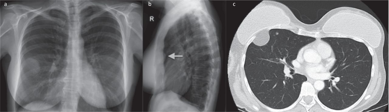
(a) AP chest radiograph showing a soft tissue opacity in the lateral aspect of the right hemithorax. (b) Lateral chest radiograph showing a soft tissue opacity posterior to the right breast. (c) CT chest scan showing a 3 cm lesion projected across the pleural margin into the right lung field. AP = anteroposterior; CT = computed tomography.

Lipomas are benign neoplastic mesenchymal tumours arising from adipose tissue. They are well defined and encapsulated. Intrathoracic lipomas are extremely rare, and usually located centrally in the anterior mediastinum, although they can also be found peripherally. They account for 1.6–2.3% of all mediastinal tumours, 0.1% of all lung tumours and 13% of all benign lung tumours.1 Most are asymptomatic and found incidentally, as in this case. However, larger, more central, lipomas may cause symptoms due to local mass effect.

The differential diagnosis of fat-containing intrathoracic tumours includes fibrolipoma, liposarcoma, teratoma, hibernoma and fibrolipomatous hamartoma.1,2 Intrathoracic lipomas can be distinguished radiologically from other tumours by the following characteristics: they are well defined, homogeneous and round, and have regular margins and a density that is similar to that of fat (approximately −100 HU).1,2 In contrast to lipomas, the other lesions listed above are not homogeneous, contain soft-tissue components and are infiltrative and much larger than lipomas; their CT density is >−50 HU.1

In most cases described in the literature, intrathoracic lipomas were surgically resected to establish the diagnosis and exclude malignancy.1,2 However, with the advancement of radiological techniques, confident diagnosis of intrathoracic lipomas and exclusion of malignancy can be made radiologically, replacing the need for invasive surgical procedures or even percutaneous biopsies.

  • © 2013 Royal College of Physicians

References

  1. ↵
    1. Yıldırım M,
    2. Parlak E,
    3. Köroglu M,
    4. et al.
    Diagnostics of peripherally located intrathoracic lipoma. Acta Inform Med 2012;20:129–30.
    OpenUrlPubMed
  2. ↵
    1. Wood J,
    2. Henderson RG
    . Peripheral intrapulmonary lipoma: a rare lung neoplasm. Br J Radiol 2004;77:60–2. doi:10.1259/bjr/94129286 doi:10.1259/bjr/94129286
    OpenUrlAbstract/FREE Full Text
Back to top
Previous articleNext article

Article Tools

Download PDF
Article Alerts
Sign In to Email Alerts with your Email Address
Citation Tools
Intrathoracic chest wall lipoma: mimicking a soft tissue neoplasm on a chest radiograph
Yasser Madani, Zara Oozeerally, Imran Syed, Akeel Jubber
Clinical Medicine Dec 2013, 13 (6) 628; DOI: 10.7861/clinmedicine.13-6-628

Citation Manager Formats

  • BibTeX
  • Bookends
  • EasyBib
  • EndNote (tagged)
  • EndNote 8 (xml)
  • Medlars
  • Mendeley
  • Papers
  • RefWorks Tagged
  • Ref Manager
  • RIS
  • Zotero
Share
Intrathoracic chest wall lipoma: mimicking a soft tissue neoplasm on a chest radiograph
Yasser Madani, Zara Oozeerally, Imran Syed, Akeel Jubber
Clinical Medicine Dec 2013, 13 (6) 628; DOI: 10.7861/clinmedicine.13-6-628
del.icio.us logo Digg logo Reddit logo Twitter logo CiteULike logo Facebook logo Google logo Mendeley logo
  • Tweet Widget
  • Facebook Like
  • Google Plus One

Jump to section

  • Article
    • References
  • Figures & Data
  • Info & Metrics

Related Articles

  • No related articles found.
  • PubMed
  • Google Scholar

Cited By...

  • No citing articles found.
  • Google Scholar

More in this TOC Section

  • Images of the month: The conundrum of chronic coccidioidomycosis
  • Images of the month: Pulmonary cement embolism
  • Images of the month: Gliptin-induced bullous pemphigoid
Show more Image of the month

Similar Articles

Navigate this Journal

  • Journal Home
  • Current Issue
  • Ahead of Print
  • Archive

Related Links

  • ClinMed - Home
  • FHJ - Home
clinmedicine Footer Logo
  • Home
  • Journals
  • Contact us
  • Advertise
HighWire Press, Inc.

Follow Us:

  • Follow HighWire Origins on Twitter
  • Visit HighWire Origins on Facebook

Copyright © 2021 by the Royal College of Physicians