
ABSTRACT
With the advent of multi-detector computed tomography, the identification of solitary pulmonary nodules is becoming ever more common. Although the prevalence of malignancy in a high risk population is only 1–1.5%, accurate identification of malignant nodules is essential to allow optimal treatment. In this article we review the most common causes of solitary pulmonary nodules and discuss diagnostic algorithms as well as some of the novel diagnostic imaging techniques in development.
Background
The incidental finding of a solitary pulmonary nodule (SPN) is taking increasing prominence in the workload of both radiologists and respiratory physicians. The introduction of helical computed tomography (CT) during the 1990s and, more recently, multidetector row CT, which can generate 1–2-mm thick slices, has enabled the identification of ever smaller subcentimetre nodules. Prevalence rates of pulmonary nodules have been reported mainly in the context of lung cancer-screening trials and vary widely from 8% to 51%,1,2 depending upon the population studied. In addition, approximately 10% of individuals will develop a new nodule over a 1-year period.3 Whereas up to half of all smokers over the age of 50 years will have at least one nodule,4 the prevalence of lung cancer in a high-risk population is only approximately 1–1.5%.2
Lung cancer is the leading cause of cancer deaths worldwide.2 The overall 5-year survival rate of patients with lung cancer across Europe is only 10% because of the large percentage of patients presenting with locally advanced or metastatic disease.2 This contrasts sharply with the good surgical outcomes for stage 1a (tumour size <2 cm) non-small cell lung cancer with postoperative 5-year survival approaching 80%.1,5 Therefore, balancing the need for identifying an early-stage lung cancer with the risk of over-investigation and/or -radiation exposure and provoking undue anxiety in patients with benign lesions is pivotal in the diagnostic evaluation and management of the SPN.
Definition
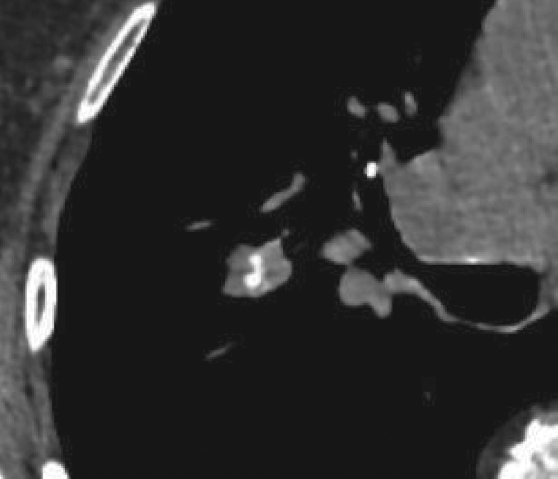
A pulmonary nodule is a round or oval lesion, 3 cm or less in diameter, of soft-tissue density that is completely surrounded by, and obliterates, the underlying lung parenchyma.6 The 3-cm cut off is arbitrary because lung nodules were originally described on chest radiographs as pulmonary opacities 1–3 cm in diameter.6,7 The advent of thin-slice spiral CT has allowed more accurate measurement of nodule size and characterisation of morphology, in particular the identification of ground-glass opacities. These are intrapulmonary nodules, with a solid component of <5% of the nodule volume, that do not attenuate the architecture of the underlying lung parenchyma.8 The term ‘subsolid nodule’ is interchangeable with ground-glass nodule (GGN) when the latter encompasses discrete pulmonary nodules that are either of pure ground-glass or have part-solid attenuation (Fig 1).8

Part-solid, part ground-glass nodule.
Differential diagnosis
The differential diagnosis of an incidental SPN encompasses up to 80 different conditions.7 The probable diagnosis in any given scenario is influenced by the individual's age, smoking history, history of occupational exposures and other risk factors for malignancy, as well as country of residence.7 Some of the infectious, non-infectious and neoplastic causes of an SPN are listed in Table 1. In the absence of any other CT features, the likelihood of a SPN being a metastasis from an extrapulmonary malignancy rather than a bronchogenic carcinoma varies according to the site of the original primary.9,10 It is more likely in cases of melanoma, sarcoma or testicular carcinoma, but less likely if the primary is a head and neck cancer, or originating from the upper gastrointestinal tract, bladder, cervix or prostate.9
Causes of a solitary pulmonary nodule.
Nodule morphology and growth as a predictor of aetiology
The SPN is often an incidental finding in an asymptomatic individual. Most malignant nodules have doubling times of approximately 21–400 days.6 Faster volume-doubling times of <20 days or slower growth rates of >400 days are more likely to reflect infectious or benign aetiologies, respectively.9 Stability of growth over 2 years implies a doubling time of more than 730 days;6 it was previous conventional wisdom that this stability reflected a benign aetiology.6,10 It is now recognised that GGNs might reflect slow-growing adenocarcinoma and their stability over 2 years reduces, but does not eliminate, the likelihood of malignancy.9
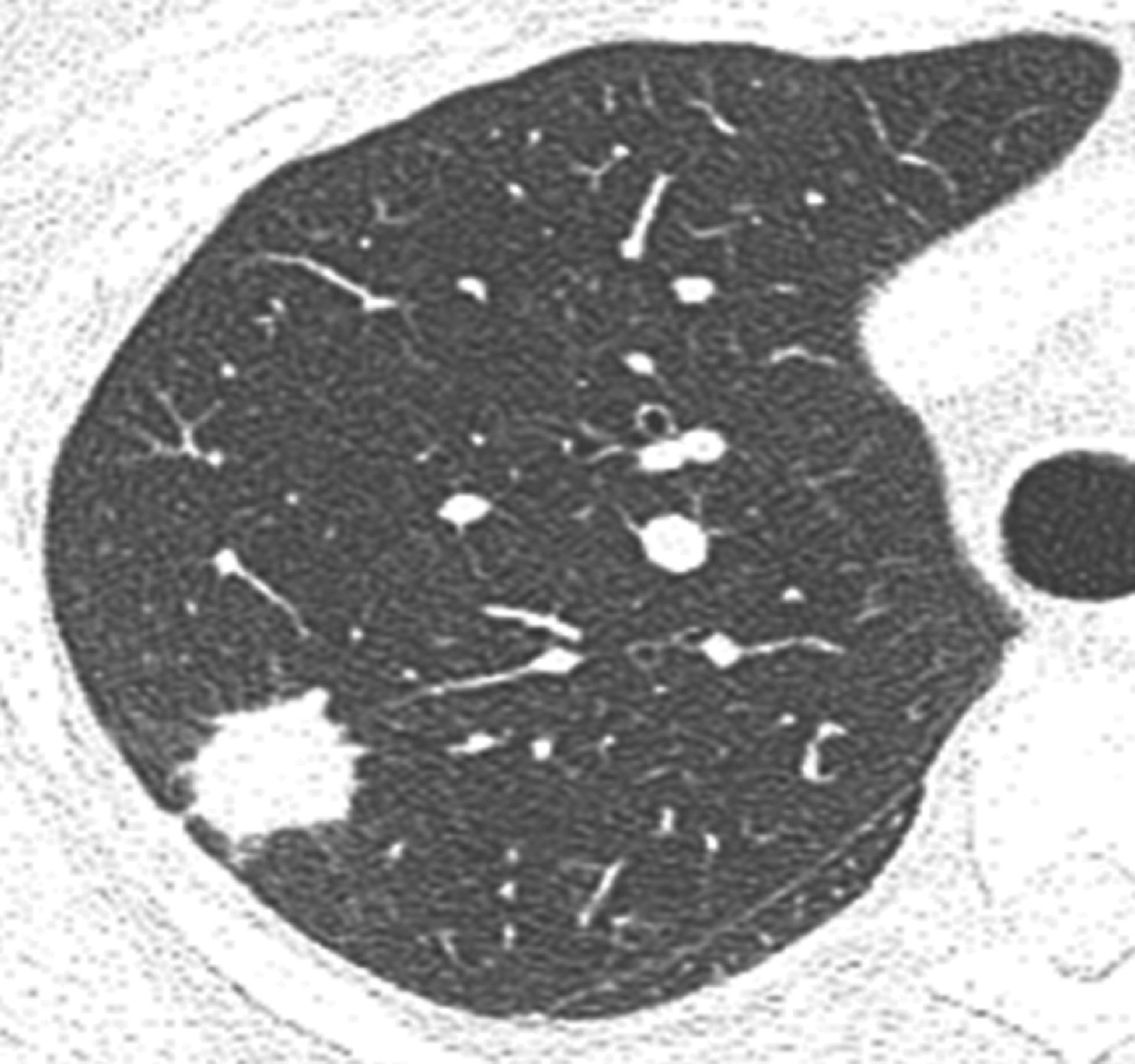
The best predictors for a nodule being benign are chronicity and patterns of calcification.10 Previous imaging, if available, might provide valuable information about chronicity and aetiology. Hamartomas can exhibit chondroid or ‘popcorn-like’ calcification (Fig 2). A wholly calcified nodule is likely to be an old granuloma, as are nodules with central or laminated calcification patterns.6 By contrast, eccentric or peripheral calcification can imply a carcinoma that has engulfed an adjacent calcified granuloma and this should raise suspicion of a malignant process.1
‘Popcorn’ pattern calcification within a hamartoma in an unenhanced computer tomograph scan of chest.
Other morphological characteristics, such as nodule margins, cavitation and wall thickness and the presence of satellite nodules, do not enable distinction between benign and malignant nodules.6 Spicules and lobulated margins can represent irregular interstitial fibrosis or infiltrative tumour growth (Fig 3).9,11 Although the presence of lobulated or spiculated margins increases the likelihood of malignancy, it is not a prerequisite, because one in five nodules with smooth margins is malignant.1,6 It is now recognised that some indeterminant subpleural or intraparenchymal nodules represent intrapulmonary lymph nodes (IPLNs).12 Ranging in size between 3 and 10 mm, IPLNs can wax and wane in size, confusing the unwary and leading to repeat CT for an otherwise ‘normal’ structure. Confident diagnosis by an experienced thoracic radiologist, based on location and morphology, can often avoid unnecessary patient anxiety.
Spiculated pulmonary nodule that is highly suggestive of malignancy.
Evaluating the growth rate of an SPN can be difficult and is dependent upon its size. For a spherical nodule, a doubling of its volume results in only a 26–30% increase in its diameter.8,10 Accurately and reproducibly measuring a 1-mm increase in diameter of a 4-mm nodule is harder than for a 3-cm mass. A 1-mm increase in diameter of a 4-mm nodule represents a doubling in volume1 that, if occurring during a certain time frame, increases the likelihood of malignancy. However, this can be easily overlooked given intra- and inter-observer variation in nodule size measurements, particularly in the presence of spiculated margins.
In the context of lung-screening trials, up to one in five positive-screening CT scans at baseline were the result of part-solid or non-solid GGNs.8 Such lesions are associated with higher rates of malignancy, with adenocarcinoma being the most common subtype.8,11 The previously used term ‘bronchioloalveolar carcinoma’ has recently been reclassified in a continuum of pre-invasive to overtly malignant subtypes, with atypical adenomatous hyperplasia being a premalignant subtype and adenocarcinoma in situ and minimally invasive adenocarcinoma describing lesions with lepidic growth patterns, with and without invasive features, respectively.13 Invasive adenocarcinoma is further subdivided according to growth patterns.13
Risk assessment
The diagnostic approach to the incidental SPN should include a thorough assessment of the individual with a full history and physical examination, with particular attention paid to factors increasing cancer risk. In addition to routinely identified risk factors for lung cancer, such as age, active and passive smoking history and occupational exposure to carcinogens, including asbestos, other risk factors should not be overlooked. Previous history of cancer of any type as well as exposure to radiotherapy, cancer in a first-degree relative and lung parenchymal abnormalities, such as chronic obstructive pulmonary disease and interstitial pulmonary fibrosis, should be recorded.
The probability of a nodule being malignant increases with age, being <3% in under-40-year olds, but 50% in those over 60 years of age.1 Furthermore, the prevalence of malignancy increases with nodule size and is estimated from lung cancer-screening study data to be <1% in nodules <5 mm in diameter, and >80% in those larger than 2 cm.1 Hence, evaluating an SPN in the context of a patient's age and other risk factors, along with its size, enables a quanitative assessment of lung cancer risk and guides the management approach.
Quantitative risk assessment of lung cancer was originally proposed during the 1970s, using Bayesian analysis,7 and has been advocated again recently.14 Various mathematical models exist using a combination of the individual's characteristics (eg age, smoking history and previous history of cancer) and features of the SPN (eg nodule size and location, and presence of spiculations) to compute a likelihood ratio of cancer.14,15 Patel et al argue that a pre-test probability calculation improves risk stratification and facilitates further management along Fleischner Society guidelines.14
Diagnostic algorithm
CT is the modality of choice for the initial diagnosis and followup of pulmonary nodules. Contrast-enhanced CT enables identification of parenchymal abnormalities as well as staging of the mediastinum and extrapulmonary structures. Unenhanced low-dose thin-slice multidetector row CT is best utilised for characterising nodule morphology and assessing growth on followup.
Positron emission tomography (PET) uses a positron-emitting radionuclide isotope of fluorine attached to a glucose analogue (18F-fluorodeoxyglucose [FDG]) that is avidly taken up by tissue with high metabolic activity. Combining PET with CT enables better anatomical correlation of parenchymal abnormality on CT with areas of high metabolic activity on PET, as well as assessment of the mediastinum for the purposes of staging. Sensitivity and specificity values of combined PET-CT have been shown by several studies to be superior to those for PET or CT alone, and have been reported to be as high as 96% and 88%, respectively, with a negative predictive value of 92%.9 FDG uptake is influenced by tumour size and metabolic activity and, thus, PET has a limited role in the diagnostic assessment of small (<8–10 mm) or purely GGNs.9,16
The approach to the management of the incidental solid SPN should follow Fleischner Society guidelines,4 outlined in Table 2. These propose surveillance CT scan followup at set intervals based on nodule size (mean of length and width) and the individual's risk factors for malignancy; the low-risk group being those with no or minimal smoking history and no risk factors for malignancy. The guidelines are not applicable to those suspected of having infection, undergoing CT as part of staging for known or suspected carcinoma of any site, or those under the age of 35 years.
Fleischner Society guidelines for CT surveillance of incidental solitary pulmonary nodules.
Fleischner Society recommendations have recently been produced for the management of subsolid nodules.13 All individuals are considered equally without risk stratification and subsolid nodules are assessed depending on their type and multiplicity. When assessing multiple nodules, the recommendations are based on the features of the dominant lesion. It is worth noting that these recommendations are based on low-dose thin-slice (1-mm) CT scans reconstructed with appropriate windows for the solid and non-solid components, with measurements performed across two dimensions and growth assessed against the original baseline CT. Pure GGNs ≤5 mm do not require followup if solitary, provided it is confirmed that they have no solid component on thin-slice CT. If multiple, interval scans at 2 and 4 years are recommended, as well as investigations to rule out other causes of multiple GGNs. If >5 mm, pure GGNs, whether solitary or multiple, require an initial scan at 3 months to confirm persistence and then annual CT scans for a minimum of 3 years. Solitary part-solid GGNs require initial CT scans at 3 months and, if persistent and with a solid component of <5 mm, can be followed annually. Part-solid GGNs with a solid component >5 mm should be considered for biopsy or surgical resection.
Novel diagnostic imaging techniques
Several imaging techniques to better enable differentiation between benign and malignant lesions are being evaluated. Nodule growth can be estimated from volumetric measurements and prediction about likelihood of malignancy inferred from calculation of volume-doubling time. Using a volume-doubling time of 500 days as the upper limit for malignancy, but excluding GGNs, Revel et al proposed a strategy to differentiate benign from malignant nodules in a retrospective case series with a sensitivity and specificity of 91% and 90%, respectively.17 Nodule volume measurements and volume-doubling time estimation using CT volumetric software is currently being utilised in the UK Lung Screen (UKLS) and the Dutch-Belgian (NELSON) lung cancer screening trials, comparing low-dose CT with usual care in those at higher risk of lung cancer.18,19
The degree of contrast enhancement of a nodule depends upon its vascularity, with correlation between nodule vascular endothelial growth factor expression and microvessel density and uptake of intravenous contrast medium.6,20 Administration of contrast followed by serial image acquisition at 60-s intervals for 4 min enables the measurement of nodule enhancement at fixed time intervals and the calculation of net enhancement values. A nodule demonstrating an increase of 15 Hounsfield units (HU) or less after injection of contrast is likely to be benign, whereas an increase of >30 HU suggests malignancy.21 A cut-off value of 15 HU has a high sensitivity (98%), reducing false negative rates, but limited specificity (58%), with an overlap in enhancement patterns and attenuation values between benign and malignant lesions. The utility of dynamic contrast-enhanced CT is being investigated in the current UK-based Solitary Pulmonary Nodule Investigation trial (SPUtNIk).22 This study also includes health economic modelling for the evaluation of SPNs.
Dynamic contrast-enhanced magnetic resonance imaging (MRI) is emerging as a tool with better sensitivity and specificity than CT or PET in distinguishing malignant from benign nodules, in particular those associated with active inflammation.9,23 Investigators have studied different aspects of this technique, with some looking at the initial first pass of contrast via the arterial system, whereas others have analysed the arterial inflow and subsequent diffusion and redistribution of contrast into the interstitial space. Distinct patterns of contrast uptake and washout demonstrated in malignant and benign inflammatory nodules have been attributed to differences in nodule microvasculature architecture.23
Summary
The SPN poses a diagnostic challenge that requires careful clinical–radiological correlation by an expert multidisciplinary team to avoid over-investigation of benign lesions, while improving lung cancer survival through detection of early-stage disease amenable to surgical resection. At present, the Fleischner guidelines form the backbone for nodule management, although novel imaging techniques might enable further refinement to this approach in the future.
- © 2013 Royal College of Physicians
References


Jump to section
Related Articles
Cited By...
- No citing articles found.