
Main menu

  • Home
  • Our journals
    • Clinical Medicine
    • Future Healthcare Journal
  • Subject collections
  • About the RCP
  • Contact us

Clinical Medicine Journal

  • ClinMed Home
  • Content
    • Current
    • Ahead of print
    • Archive
  • Author guidance
    • Instructions for authors
    • Submit online
  • About ClinMed
    • Scope
    • Editorial board
    • Policies
    • Information for reviewers
    • Advertising

User menu

  • Log in

Search

  • Advanced search
RCP Journals
Home
  • Log in
  • Home
  • Our journals
    • Clinical Medicine
    • Future Healthcare Journal
  • Subject collections
  • About the RCP
  • Contact us
Advanced

Clinical Medicine Journal

clinmedicine Logo
  • ClinMed Home
  • Content
    • Current
    • Ahead of print
    • Archive
  • Author guidance
    • Instructions for authors
    • Submit online
  • About ClinMed
    • Scope
    • Editorial board
    • Policies
    • Information for reviewers
    • Advertising

Vanishing lung disease in an adult misdiagnosed as pneumothorax

Abhishek Jha, Ajay Gupta, Prakhar Gupta, Gagan Gupta, Ibne Ahmad and Mohd Saud
Download PDF
DOI: https://doi.org/10.7861/clinmedicine.14-2-210
Clin Med April 2014
Abhishek Jha
ADepartment of Radiodiagnosis, Jawaharlal Nehru Medical College, Aligarh Muslim University, Aligarh, Uttar Pradesh, India
Roles: senior resident
  • Find this author on Google Scholar
  • Find this author on PubMed
  • Search for this author on this site
  • For correspondence: abhi.jnmc@gmail.com
Ajay Gupta
BDepartment of Pharmacology, Jawaharlal Nehru Medical College, Aligarh Muslim University, Aligarh, Uttar Pradesh, India
Roles: junior resident
  • Find this author on Google Scholar
  • Find this author on PubMed
  • Search for this author on this site
Prakhar Gupta
CDepartment of General Surgery, Jawaharlal Nehru Medical College, Aligarh Muslim University, Aligarh, Uttar Pradesh, India
Roles: junior resident
  • Find this author on Google Scholar
  • Find this author on PubMed
  • Search for this author on this site
Gagan Gupta
DDepartment of Radiodiagnosis, Jawaharlal Nehru Medical College, Aligarh Muslim University, Aligarh, Uttar Pradesh, India
Roles: senior resident
  • Find this author on Google Scholar
  • Find this author on PubMed
  • Search for this author on this site
Ibne Ahmad
EDepartment of Radiodiagnosis, Jawaharlal Nehru Medical College, Aligarh Muslim University, Aligarh, Uttar Pradesh, India
Roles: professor
  • Find this author on Google Scholar
  • Find this author on PubMed
  • Search for this author on this site
Mohd Saud
FDepartment of Radiodiagnosis, Jawaharlal Nehru Medical College, Aligarh Muslim University, Aligarh, Uttar Pradesh, India
Roles: junior resident
  • Find this author on Google Scholar
  • Find this author on PubMed
  • Search for this author on this site
  • Article
  • Figures & Data
  • Info & Metrics
Loading

A previously healthy 34-year-old man attended the medical outpatient ward with a 6-month history of shortness of breath, which was worse on exertion. The patient was a chronic smoker but there was no additional history of cough, sputum production, haemoptysis, fever or chest pain. On examination, the patient appeared pallid and had reduced tactile vocal fremitus and breath sounds bilaterally.

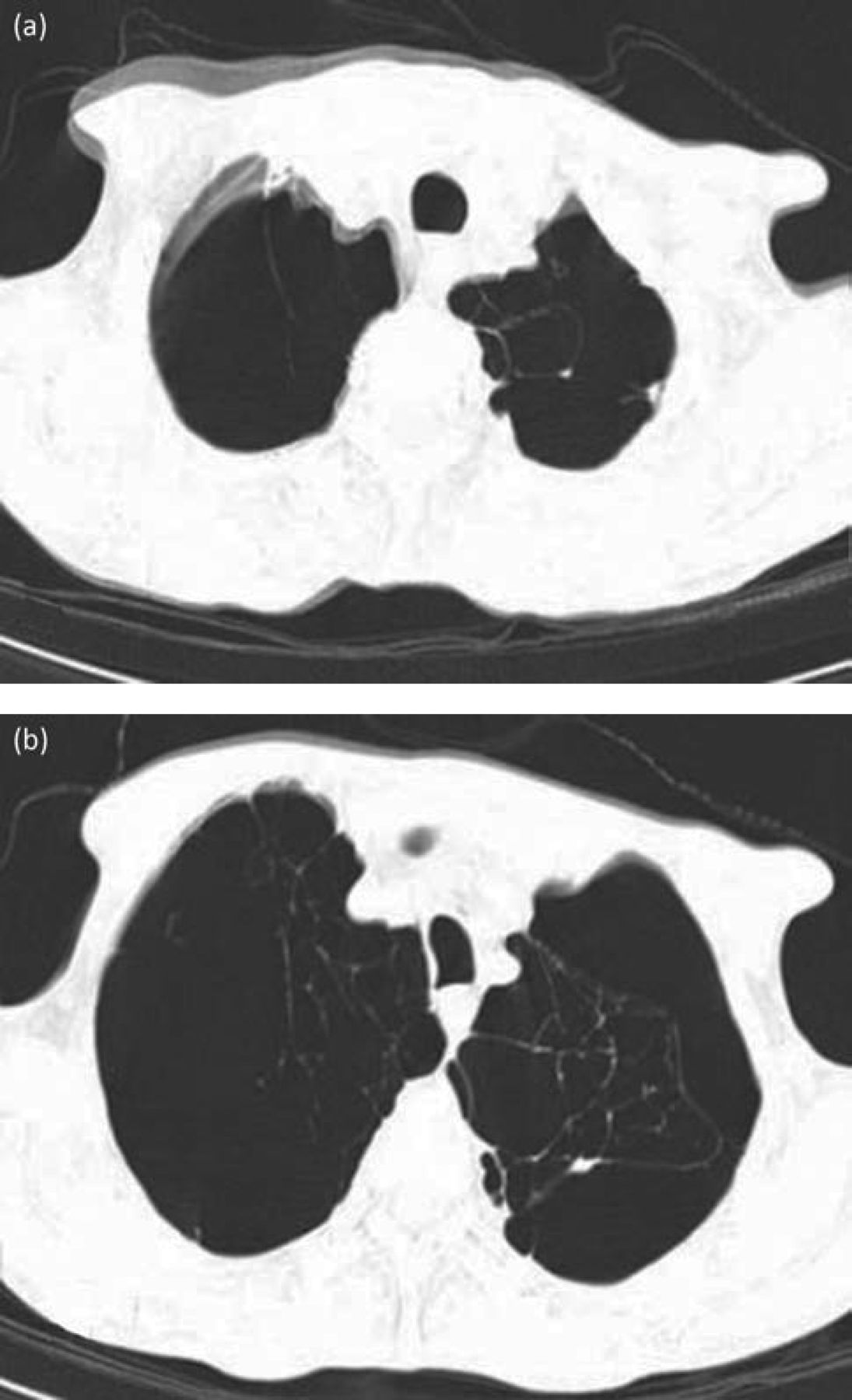
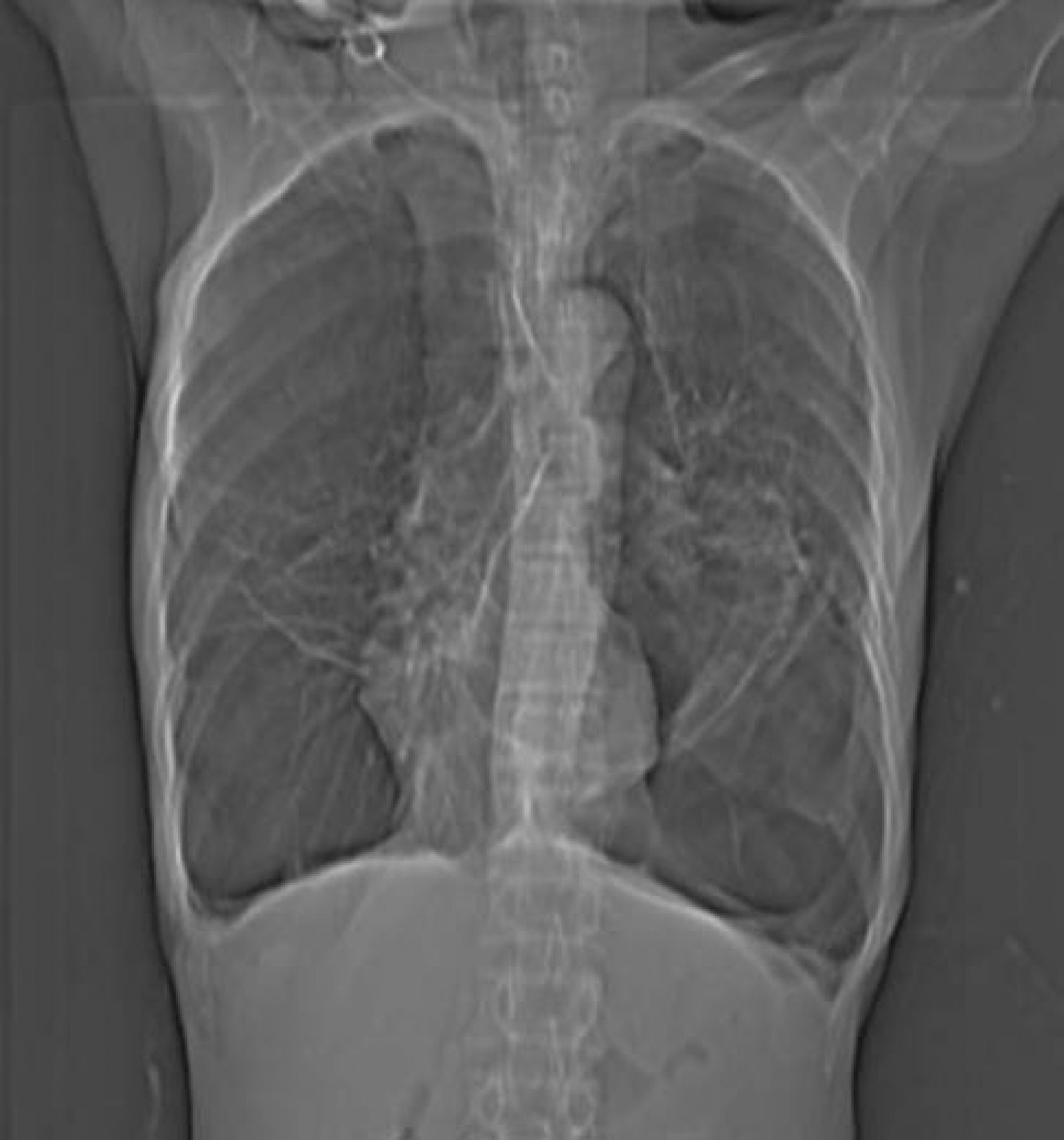
A plain chest X-ray revealed multiple bullae bilaterally (Fig 1). There was no tracheal or mediastinal displacement. A diagnosis of pneumothorax was considered and a tube thoracostomy was planned. A high resolution chest computed tomography (HRCT) scan was organised to confirm the diagnosis. However, this revealed multiple large bullae in bilateral lung fields with gross destruction of the pulmonary architecture (Fig 2 and 3). Pulmonary function tests demonstrated reduced lung volume and the presence of a restrictive deficit.

The diagnosis in this case was vanishing lung syndrome, first described by Burke et al in 1937.1 It is characterised by unilateral or bilateral asymmetric upper lobe involvement with the formation of multiple bullae. It is most commonly seen in young male smokers who present with progressive breathlessness. The pathogenesis of the disease revolves around the destruction of alveolar walls resulting in the formation of sub-pleural plebs with coalesce to create giant bullae and compression of normal lung tissue. The disease follows a downhill course, leading to respiratory failure and eventual death. It can be challenging to differentiate from pneumothorax, which imparts a similar appearance on plain chest X-ray. The ‘double wall sign’ seen in pneumothorax is due to air along both sides of the bullae and can be highly useful in distinguishing between the two entities.2

Treatment of the disease depends on the clinical picture. In the early stages, smoking cessation is important along with supportive management. Advanced cases require surgery. For our patient, a pneumonectomy was recommended, but the patient was not keen to proceed and was thus managed conservatively.

Fig 1.
  • Download figure
  • Open in new tab
  • Download powerpoint
Fig 1.

Axial HRCT of the chest through upper lung zones shows multiple bullae replacing the pulmonary architecture. HRCT = high resolution chest computed tomography.

Fig 2.
  • Download figure
  • Open in new tab
  • Download powerpoint
Fig 2.

Axial HRCT of the chest through lower lung zones showing giant bullae occupying more than one-third of hemithorax in bilateral lung bases. HRCT = high resolution chest computed tomography.

Fig 3.
  • Download figure
  • Open in new tab
  • Download powerpoint
Fig 3.

Frontal chest radiograph showing multiple bullae in bilateral lung fields. However, the possibility of pneumothorax cannot be excluded.

  • © 2014 Royal College of Physicians

References

  1. ↵
    1. Burke R
    . Vanishing lungs: a case report of bullous emphysema. Radiology 1937;28:367–71.
    OpenUrlCrossRef
  2. ↵
    1. Phillips GD,
    2. Trotman-Dickensen B,
    3. Hodson ME,
    4. Geddes DM
    . Role of CT in the management of pneumothorax in patients with complex cystic lung disease. Chest 1997;112:275–8.doi:10.1378/chest.112.1.275
    OpenUrlCrossRefPubMed
Back to top
Previous articleNext article

Article Tools

Download PDF
Article Alerts
Sign In to Email Alerts with your Email Address
Citation Tools
Vanishing lung disease in an adult misdiagnosed as pneumothorax
Abhishek Jha, Ajay Gupta, Prakhar Gupta, Gagan Gupta, Ibne Ahmad, Mohd Saud
Clinical Medicine Apr 2014, 14 (2) 210-211; DOI: 10.7861/clinmedicine.14-2-210

Citation Manager Formats

  • BibTeX
  • Bookends
  • EasyBib
  • EndNote (tagged)
  • EndNote 8 (xml)
  • Medlars
  • Mendeley
  • Papers
  • RefWorks Tagged
  • Ref Manager
  • RIS
  • Zotero
Share
Vanishing lung disease in an adult misdiagnosed as pneumothorax
Abhishek Jha, Ajay Gupta, Prakhar Gupta, Gagan Gupta, Ibne Ahmad, Mohd Saud
Clinical Medicine Apr 2014, 14 (2) 210-211; DOI: 10.7861/clinmedicine.14-2-210
Reddit logo Twitter logo Facebook logo Mendeley logo
  • Tweet Widget
  • Facebook Like
  • Google Plus One

Jump to section

  • Article
    • References
  • Figures & Data
  • Info & Metrics

Related Articles

  • No related articles found.
  • PubMed
  • Google Scholar

Cited By...

  • No citing articles found.
  • Google Scholar

More in this TOC Section

  • Purple urine bag syndrome
  • Bullous Sweet's syndrome in rheumatoid arthritis after streptococcal pharyngitis
  • A distinctive posterior mitral valve infective endocarditis and a large mycotic aneurysm
Show more Image of the month

Similar Articles

FAQs

  • Difficulty logging in.

There is currently no login required to access the journals. Please go to the home page and simply click on the edition that you wish to read. If you are still unable to access the content you require, please let us know through the 'Contact us' page.

  • Can't find the CME questionnaire.

The read-only self-assessment questionnaire (SAQ) can be found after the CME section in each edition of Clinical Medicine. RCP members and fellows (using their login details for the main RCP website) are able to access the full SAQ with answers and are awarded 2 CPD points upon successful (8/10) completion from:  https://cme.rcplondon.ac.uk

Navigate this Journal

  • Journal Home
  • Current Issue
  • Ahead of Print
  • Archive

Related Links

  • ClinMed - Home
  • FHJ - Home
clinmedicine Footer Logo
  • Home
  • Journals
  • Contact us
  • Advertise
HighWire Press, Inc.

Follow Us:

  • Follow HighWire Origins on Twitter
  • Visit HighWire Origins on Facebook

Copyright © 2021 by the Royal College of Physicians