
Main menu

  • Home
  • Our journals
    • Clinical Medicine
    • Future Healthcare Journal
  • Subject collections
  • About the RCP
  • Contact us

Clinical Medicine Journal

  • ClinMed Home
  • Content
    • Current
    • Ahead of print
    • Archive
  • Author guidance
    • Instructions for authors
    • Submit online
  • About ClinMed
    • Scope
    • Editorial board
    • Policies
    • Information for reviewers
    • Advertising

User menu

  • Log in

Search

  • Advanced search
RCP Journals
Home
  • Log in
  • Home
  • Our journals
    • Clinical Medicine
    • Future Healthcare Journal
  • Subject collections
  • About the RCP
  • Contact us
Advanced

Clinical Medicine Journal

clinmedicine Logo
  • ClinMed Home
  • Content
    • Current
    • Ahead of print
    • Archive
  • Author guidance
    • Instructions for authors
    • Submit online
  • About ClinMed
    • Scope
    • Editorial board
    • Policies
    • Information for reviewers
    • Advertising

Point-of-care cardiac ultrasound in acute medicine – the quick scan

Sandeep S. Hothi, David Sprigings and John Chambers
Download PDF
DOI: https://doi.org/10.7861/clinmedicine.14-6-608
Clin Med December 2014
Sandeep S. Hothi
AMurray Edwards College, University of Cambridge, Cambridge, UK, and clinical lecturer in cardiology, Glenfield Hospital, Leicester, UK
Roles: fellow
  • Find this author on Google Scholar
  • Find this author on PubMed
  • Search for this author on this site
  • For correspondence: ssh26@cam.ac.uk
David Sprigings
BNorthampton General Hospital, Northampton, UK
Roles: consultant cardiologist
  • Find this author on Google Scholar
  • Find this author on PubMed
  • Search for this author on this site
John Chambers
CGuy's and St Thomas’ Hospital, London, UK
Roles: consultant cardiologist and professor of clinical cardiology
  • Find this author on Google Scholar
  • Find this author on PubMed
  • Search for this author on this site
  • Article
  • Figures & Data
  • Info & Metrics
Loading

ABSTRACT

The optimum management of acute medical patients requires prompt and accurate diagnosis, monitoring and treatment. The clinical history and physical examination remain central to diagnosis, but often need supplementation by laboratory testing or imaging. Echocardiographic assessment of cardiac structure and function provides valuable information that can aid diagnosis and assess clinical progress. It has many advantages as an imaging modality, and recent technological advances have resulted in hand-held, battery-powered ultrasound devices that provide high-quality images. Three broad applications of cardiac ultrasound now exist: conventional echocardiography, focussed echocardiography and the quick-scan. A quick-scan using a hand-held ultrasound device is readily integrated into the bedside clinical assessment, providing information that can be used immediately in diagnostic reasoning; it can also guide pericardiocentesis. Hand-held ultrasound devices can also be used in acute situations, as well as geographically remote areas or special situations (eg disaster zones) where other imaging is not available. However, the diagnostic yield of echocardiography is user dependent, and training is required for its benefits to be realised, adding to the hardware costs. More data are needed on the incremental value of hand-held ultrasonography and a quick-scan over conventional methods of assessment, their impact on clinical outcomes, and cost effectiveness.

KEYWORDS
  • Quick-scan
  • point-of-care
  • acute medicine
  • echocardiography
  • physical examination
  • hand-held ultrasound

Introduction

Echocardiography assesses cardiac structure and function more accurately than does physical examination.1 Its use has uncovered previously unsuspected cardiac abnormalities. For example, up to 20% of patients thought to have acute chronic obstructive pulmonary disease (COPD) exacerbations might in fact have left ventricular (LV) failure.2 Thus, echocardiography is integral to acute medicine and is included within the UK acute medicine syllabus.3 Hitherto, echocardiography has usually been delivered from the cardiology department within office hours, but there is now a worldwide trend towards echocardiography training for acute, emergency and intensive care physicians, to provide point-of-care scanning. This has been facilitated by the development of portable and, more recently, hand-held ultrasound (H-USS) devices. Hand-held echocardiography (the ‘quick-scan’) is poised to become an extension of the clinical examination directly relevant to managing acute illness. This is a potentially liberating and powerful development, but there are also concerns principally relating to training and governance. Here, we discuss the applications, advantages and limitations of devolved quick-scan cardiac ultrasound by H-USS.

Hand-held ultrasound devices

Three categories of echocardiography machine exist. ‘High-end’ machines typically cost around £100,000 and are large and better suited to the echocardiography department. Portable machines typically cost £30,000–£40,000 and are often battery powered, enabling movement between beds without being turned off. They have slightly inferior image quality and less advanced software compared with high-end machines. H-USS devices costing around £5,000 are portable and ideal for integration with the clinical examination. They include the Acuson P10 (Siemens Healthcare, Surrey, UK) and VScan (GE Healthcare, Buckinghamshire, UK) (Fig 1). The latter measures 135 × 73 × 28 mm, weighs 390 g, and comprises a phased-array ultrasound probe attached by a thin cable to a rechargeable, flip-screen device with an 8.9-cm display that shows 2D images with colour Doppler.

Fig 1.
  • Download figure
  • Open in new tab
  • Download powerpoint
Fig 1.

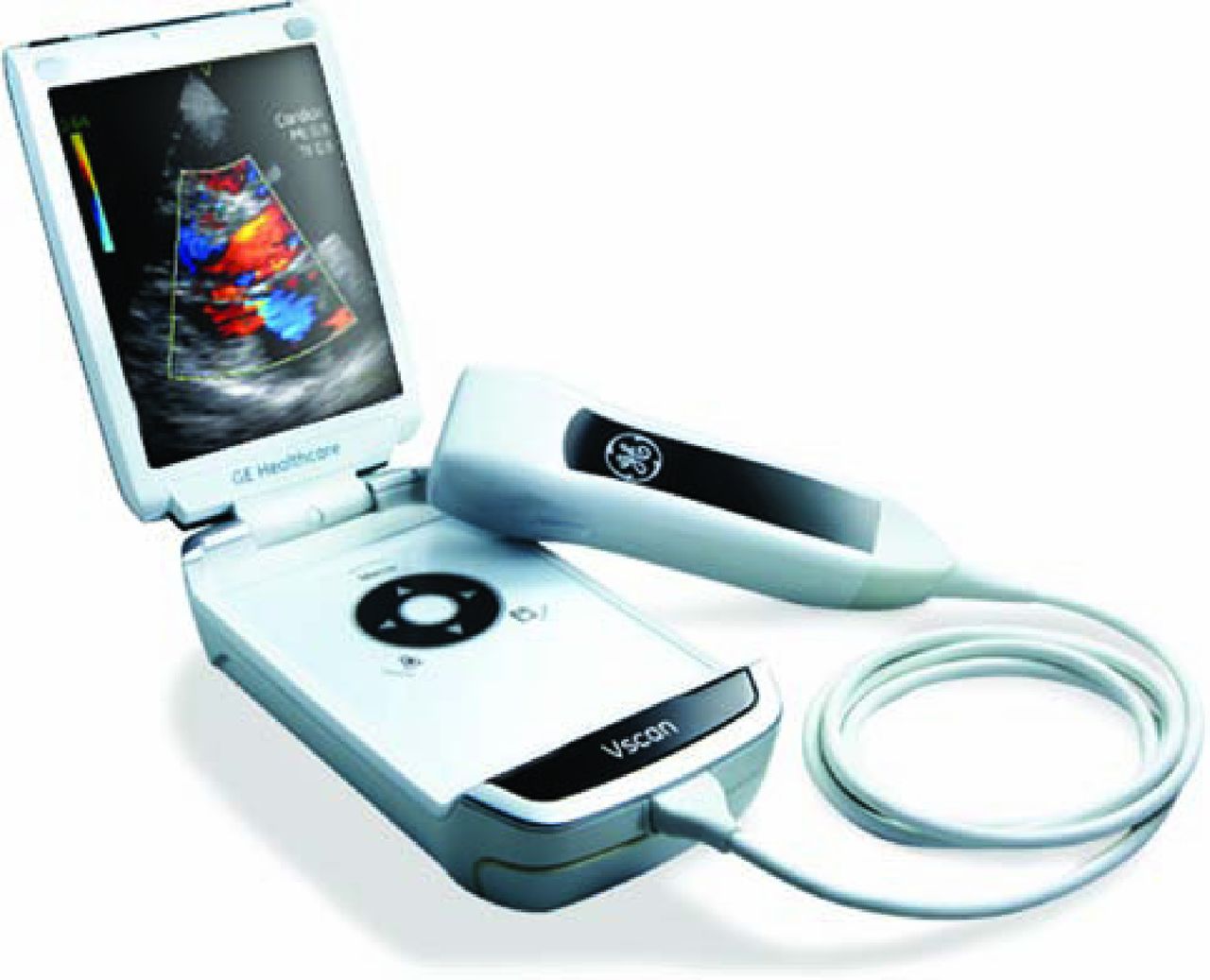
The VScan. Reproduced with permission of GE Healthcare. © 2014 General Electric Company.

Standard and portable echocardiography machines have large internal hard-drives and can easily be backed up onto electronic storage systems that enable viewing and analysis using readily available software. The hand-held machines currently have limited internal storage on a flash card, which needs to be downloaded to a PC. This is relatively time consuming and many physicians choose not to archive studies and to write a report in the patients’ case-files rather than using the departmental electronic reporting system.

These technical categories broadly correspond to three types of scan, although there can be overlap: standard echocardiography, focused echocardiography or a quick-scan (Table 1). The standard echocardiogram is the comprehensive study, typically taking 30–40 min, involving 11 views and multiple modalities. A focused study addresses a simple question, for example, assessment of residual effusion after pericardiocentesis, and is usually only safe as follow-up to a standard study. There is no consensus on the components of a quick-scan. We use it to exclude significant disease in four areas: LV size and function; right ventricular (RV) size and function; valvular structure and function; and pericardial fluid.

View this table:
  • View inline
  • View popup
  • Download powerpoint
Table 1.

Different applications of cardiac ultrasound.

Clinical applications, advantages and limitations in acute medicine of a quick-scan using a hand-held ultrasound device

There are several indications for a quick-scan (Table 2) and many benefits (Box 1). A quick-scan is rapid4 and can quickly identify pericardial effusion, severe LV systolic dysfunction (LVSD), RV dilatation, major heart valve disease and inferior vena cava (IVC) dilatation, allowing immediate potentially life-saving changes in management. There is good agreement with standard echocardiography.4–10 It improves the differentiation and management of conditions with overlapping features but contrasting treatments, such as acute LV failure versus acute exacerbation of COPD. This is additionally important where a treatment is contraindicated in the competing diagnosis, or where treatment carries great risk (eg a quick-scan could substantiate the decision to thrombolyse a moribund patient with clinically suspected pulmonary embolism). It can also guide pericardiocentesis.

View this table:
  • View inline
  • View popup
  • Download powerpoint
Table 2.

Indications for quick-scan echocardiography along with typical echocardiographic features suggestive of, and consistent with, common medical conditions.

A quick-scan is suited to acute settings, including the acute medical unit, inpatients,7,11–13 emergency department,8 cardiac arrests, intensive care,14,15 the perioperative period,5 and special situations, such as mobile medical units, disaster zones and battle zones.4,15–18 In cardiac arrest, a quick-scan can identify or indicate the presence of reversible causes of pulseless electrical activity (PEA), and distinguish true PEA, which carries a worse prognosis, from pseudo-PEA.19,20 A quick-scan can be used to triage those who need a standard echocardiogram and contributes to clinical risk stratification.

A quick-scan is not a standard echocardiogram and it has limitations. It is inferior at assessing LV dimensions,18 RV function and mitral regurgitation,18 and cannot assess diastolic function. For emergency triage, it performs well when used by operators trained in standard echocardiography, but there are few data for operators trained solely to the level of a quick-scan save for one small study in medical students.21 This has implications for who can use H-USS. Furthermore, there has been no systematic study of the impact of quick-scans on patient outcomes or costs. The European Society of Cardiology has issued a statement that H-USS should be used in a limited manner, users without accreditation in transthoracic echocardiography should receive formal training and, finally, the patient should be told that the H-USS study does not replace a full study.22

Training issues

A quick-scan can be regarded as an extension of the physical examination with the H-USS acting as an ultrasonic stethoscope. Clinicians with widely differing examination skills and clinical qualifications (eg MRCP) are all entrusted with a stethoscope. However, broad consensus requires demonstration of competency to use an ultrasound machine. This is partly because our culture expects this with the new application of technology, partly because the technique resembles standard echocardiography and partly because more is at stake. Almost no clinical decisions are acted upon without confirmation with a test, whereas the quick-scan, to be valid, should often prompt a change in management. International training programmes exist for peri-arrest quick-scans, including FAST and FEEL. However, no nationally agreed accreditation system for USS in acute medicine exists. In its absence, individual centres are free to manage their own programmes and this is one property of a British Society of Echocardiography-accredited department. One system has been piloted since 200723,24 conforming to the requirements of a recent American Society of Echocardiography consensus statement.25

Box 1. Clinical benefits of the quick-scan.


Embedded Image

The problem with accreditation is not the delivery of initial training but providing the experience for a logbook under a mentor. Furthermore, once an individual is accredited, it is vital that a devolved quick-scan service should be integrated within a total service allowing immediate back-up transthoracic echocardiography if needed, second opinions, clinical opinions, continuing professional development and quality assurance. This might require a reorganisation of echocardiography as a core hospital service rather than a subdepartment within cardiology, and echocardiographers designated for training and maintaining competence in devolved echocardiography. Finally, we propose that clinicians start training in medical school.

Potential developments in hand-held ultrasonography

H-USS devices will require secure data storage using standardised formats viewable in the more popular imaging suites to aid usability. Data should be easily archived into long-term storage systems that integrate with a patient's medical records. Alternatively, the role of imaging with H-USS needs to be agreed upon if data are not stored in a long-term system. For instance, physical examinations are not recorded digitally but the written record is taken as sufficient evidence of the examination findings. Incorporation of M-mode and various Doppler modes could improve quantification by H-USS. In the future, H-USS could use a smartphone coupled to a probe, physically or wirelessly.

Conclusion

H-USS in trained hands adds value to the physical examination and is ideally suited to acute medicine. It can be performed rapidly at the point of care and improves diagnosis, emergency decision-making and guidance of pericardiocentesis. Laennec produced the first stethoscope in 181626 and, despite early resistance from clinicians, significantly advanced the physical examination. Quick-scans using H-USS provide another potential revolution in clinical examination.27

  • © 2014 Royal College of Physicians

References

  1. 1.↵
    1. Fedson S,
    2. Neithardt G,
    3. Thomas P,
    4. et al.
    Unsuspected clinically important findings detected with a small portable ultrasound device in patients admitted to a general medical service. J Am Soc Echocardiogr 2003;16:901–5.doi:10.1016/S0894-7317(03)00426-7
    OpenUrlCrossRefPubMed
  2. 2.↵
    1. Hawkins NM,
    2. Petrie MC,
    3. Jhund PS,
    4. et al.
    Heart failure and chronic obstructive pulmonary disease: diagnostic pitfalls and epidemiology. Eur J Hear Fail 2009;11:130–9.doi:10.1093/eurjhf/hfn013
    OpenUrlCrossRef
  3. 3.↵
    1. Joint Royal Colleges of Physicians’ Training Board
    , 2014. Available online at www.jrcptb.org.uk [Accessed 5 August 2014].
  4. 4.↵
    1. Kajimoto K,
    2. Madeen K,
    3. Nakayama T,
    4. et al.
    Rapid evaluation by lung-cardiac-inferior vena cava (LCI) integrated ultrasound for differentiating heart failure from pulmonary disease as the cause of acute dyspnea in the emergency setting. Cardiovasc Ultrasound 2012;10:49.doi:10.1186/1476-7120-10-49
    OpenUrlCrossRefPubMed
  5. 5.↵
    1. Frederiksen C,
    2. Juhl-Olsen P,
    3. Larsen UT,
    4. et al.
    New pocket echocardiography device is interchangeable with high-end portable system when performed by experienced examiners. Acta Anaesthesiol Scand 2010;54:1217–23.doi:10.1111/j.1399-6576.2010.02320.x
    OpenUrlCrossRefPubMed
  6. 6.↵
    1. Réant P,
    2. Dijos M,
    3. Arsac F,
    4. et al.
    Validation of a new bedside echoscopic heart examination resulting in an improvement in echo-lab workflow. Arch Cardiovasc Dis 2011;104:171–7.doi:10.1016/j.acvd.2011.01.003
    OpenUrlCrossRefPubMed
  7. 7.↵
    1. Egan M,
    2. Ionescu A
    . The pocket echocardiograph: a useful new tool? Eur J Echocardiogr 2008;9:721–5.doi:10.1093/ejechocard/jen177
    OpenUrlAbstract/FREE Full Text
  8. 8.↵
    1. Biais M,
    2. Carrié C,
    3. Delaunay F,
    4. et al.
    Evaluation of a new pocket echoscopic device for focused cardiac ultrasonography in an emergency setting. Crit Care 2012;16:R82.doi:10.1186/cc11340
    OpenUrlCrossRefPubMed
  9. 9.↵
    1. Lafitte S,
    2. Alimazighi N,
    3. Reant P,
    4. et al.
    Validation of the smallest pocket echoscopic device's diagnostic capabilities in heart investigation. Ultrasound Med Biol 2011;37:798–804.doi:10.1016/j.ultrasmedbio.2011.02.010
    OpenUrlCrossRefPubMed
  10. 10.↵
    1. Prinz C,
    2. Voigt J-U
    . Diagnostic accuracy of a hand-held ultrasound scanner in routine patients referred for echocardiography. J Am Soc Echocardiogr 2011;24:111–6.doi:10.1016/j.echo.2010.10.017
    OpenUrlCrossRefPubMed
  11. 11.↵
    1. Galderisi M,
    2. Santoro A,
    3. Versiero M,
    4. et al.
    Improved cardiovascular diagnostic accuracy by pocket size imaging device in non-cardiologic outpatients: the NaUSiCa (Naples Ultrasound Stethoscope in Cardiology) study. Cardiovasc Ultrasound 2010;8:51.doi:10.1186/1476-7120-8-51
    OpenUrlCrossRefPubMed
  12. 12.↵
    1. Cardim N,
    2. Fernandez Golfin C,
    3. Ferreira D,
    4. et al.
    Usefulness of a new miniaturized echocardiographic system in outpatient cardiology consultations as an extension of physical examination. J Am Soc Echocardiogr 2011;24:117–24.doi:10.1016/j.echo.2010.09.017
    OpenUrlCrossRefPubMed
  13. 13.↵
    1. Liebo MJ,
    2. Israel RL,
    3. Lillie EO,
    4. et al.
    Is pocket mobile echocardiography the next-generation stethoscope? A cross-sectional comparison of rapidly acquired images with standrd transthoracic echocardiography. Ann Intern Med 2011;155:33–8.doi:10.7326/0003-4819-155-1-201107050-00005
    OpenUrlCrossRefPubMed
  14. 14.↵
    1. Vignon P,
    2. Chastagner C,
    3. François B,
    4. et al.
    Diagnostic ability of hand-held echocardiography in ventilated critically ill patients. Crit Care 2003;7:R84–91.doi:10.1186/cc2360
    OpenUrlCrossRefPubMed
  15. 15.↵
    1. Amiel J-B,
    2. Grümann A,
    3. Lhéritier G,
    4. et al.
    Assessment of left ventricular ejection fraction using an ultrasonic stethoscope in critically ill patients. Crit Care 2012;16:R29.doi:10.1186/cc11198
    OpenUrlCrossRefPubMed
  16. 16.↵
    1. Connors A,
    2. McCaffree D,
    3. Gray B
    . Evaluation of right-heart catheterization in the critically ill patient without acute myocardial infarction. N Engl J Med 1983;308:263–7.doi:10.1056/NEJM198302033080508
    OpenUrlCrossRefPubMed
  17. 17.↵
    1. Eisenberg P,
    2. Jaffe A,
    3. Schuster D
    . Clinical evaluation compared to pulmonary artery catheterization in the haemodynamic assessment of critically ill patients. Crit Care Med 1984;12:549–53.doi:10.1097/00003246-198407000-00001
    OpenUrlCrossRefPubMed
  18. 18.↵
    1. Testuz A,
    2. Müller H,
    3. Keller P-F,
    4. et al.
    Diagnostic accuracy of pocket-size handheld echocardiographs used by cardiologists in the acute care setting. Eur Heart J Cardiovasc Imaging 2013;14:38–42.doi:10.1093/ehjci/jes085
    OpenUrlAbstract/FREE Full Text
  19. 19.↵
    1. Volpicelli G
    . Usefulness of emergency ultrasound in nontraumatic cardiac arrest. Am J Emerg Med 2011;29:216–23.doi:10.1016/j.ajem.2009.03.018
    OpenUrlCrossRefPubMed
  20. 20.↵
    1. Resuscitation Council (UK)
    , 2010. Available online at www.resus.org.uk/pages/guide.htm [Accessed 5 August 2014].
  21. 21.↵
    1. Decara J,
    2. Kirkpatrick J,
    3. Spencer K,
    4. et al.
    Use of hand-carried ultrasound devices to augment the accuracy of medical student bedside cardiac diagnoses. J Am Soc Echocardiogr 2005;18:257–63.doi:10.1016/j.echo.2004.11.015
    OpenUrlCrossRefPubMed
  22. 22.↵
    1. Sicari R,
    2. Galderisi M,
    3. Voigt J-U,
    4. et al.
    The use of pocket-size imaging devices: a position statement of the European Association of Echocardiography. Eur J Echocardiogr 2011;12:85–7.doi:10.1093/ejechocard/jeq184
    OpenUrlAbstract/FREE Full Text
  23. 23.↵
    1. Victor K,
    2. Rajani R,
    3. Bruemmer-Smith S,
    4. Kabir S,
    5. Chambers J
    . A training programme in screening echocardiography. Clin Teach 2013;10:176–80.doi:10.1111/tct.12019
    OpenUrlCrossRefPubMed
  24. 24.↵
    1. Chambers J,
    2. Bruemmer-Smith S,
    3. Hindocha R,
    4. Langrish,
    5. Johnson A
    . Basic screening echocardiography: a training programme. J Int Care Soc 2011;12:2–3.
    OpenUrl
  25. 25.↵
    1. Spencer K,
    2. Kimura B,
    3. Korcarz C,
    4. et al.
    Focused cardiac ultrasound: recommendations from the American Society of Echocardiography. J Am Soc Echocardiogr 2013;26:567–81.doi:10.1016/j.echo.2013.04.001
    OpenUrlCrossRefPubMed
  26. 26.↵
    1. Laennec R.
    De l’auscultation médiate ou Traité du Diagnostic des Maladies des Poumon et du Coeur. Paris: Brosson & Chaudé; 1819.
  27. 27.↵
    1. Solomon S,
    2. Saldana F
    . Point-of-care ultrasound in medical education – stop listening and look. N Engl J Med 2014;370:1083–5.doi:10.1056/NEJMp1311944
    OpenUrlCrossRefPubMed
View Abstract
Back to top
Previous articleNext article

Article Tools

Download PDF
Article Alerts
Sign In to Email Alerts with your Email Address
Citation Tools
Point-of-care cardiac ultrasound in acute medicine – the quick scan
Sandeep S. Hothi, David Sprigings, John Chambers
Clinical Medicine Dec 2014, 14 (6) 608-611; DOI: 10.7861/clinmedicine.14-6-608

Citation Manager Formats

  • BibTeX
  • Bookends
  • EasyBib
  • EndNote (tagged)
  • EndNote 8 (xml)
  • Medlars
  • Mendeley
  • Papers
  • RefWorks Tagged
  • Ref Manager
  • RIS
  • Zotero
Share
Point-of-care cardiac ultrasound in acute medicine – the quick scan
Sandeep S. Hothi, David Sprigings, John Chambers
Clinical Medicine Dec 2014, 14 (6) 608-611; DOI: 10.7861/clinmedicine.14-6-608
del.icio.us logo Digg logo Reddit logo Twitter logo CiteULike logo Facebook logo Google logo Mendeley logo
  • Tweet Widget
  • Facebook Like
  • Google Plus One

Jump to section

  • Article
    • ABSTRACT
    • Introduction
    • Hand-held ultrasound devices
    • Clinical applications, advantages and limitations in acute medicine of a quick-scan using a hand-held ultrasound device
    • Training issues
    • Potential developments in hand-held ultrasonography
    • Conclusion
    • References
  • Figures & Data
  • Info & Metrics

Related Articles

  • No related articles found.
  • PubMed
  • Google Scholar

Cited By...

  • Murmur clinic: validation of a new model for detecting heart valve disease
  • Detecting heart valve disease: can we do better?
  • Google Scholar

More in this TOC Section

  • Management of high-risk non-ST elevation myocardial infarction in the UK: need for alternative models of care to reduce length of stay and admission to angiography times
  • Transcatheter aortic valve implantation – what the general physician needs to know
  • Diabetes patient at risk score – a novel system for triaging appropriate referrals of inpatients with diabetes to the diabetes team
Show more Clinical Practice

Similar Articles

Navigate this Journal

  • Journal Home
  • Current Issue
  • Ahead of Print
  • Archive

Related Links

  • ClinMed - Home
  • FHJ - Home
clinmedicine Footer Logo
  • Home
  • Journals
  • Contact us
  • Advertise
HighWire Press, Inc.

Follow Us:

  • Follow HighWire Origins on Twitter
  • Visit HighWire Origins on Facebook

Copyright © 2020 by the Royal College of Physicians