
A 55-year-old male presented with progressive dyspnoea for five years. He had worked in the marble industry for the past 20 years and was a stone crusher. Chest radiograph revealed bilateral radiodense masses in a parahilar location (Fig 1). On CT, the soft tissue masses were of high attenuation with emphysematous changes in adjacent lung. The lateral margins of the mass paralleled the lateral chest wall. In addition, multiple small centrilobular nodules were noted in bilateral upper lobes (Fig 2). Few enlarged mediastinal lymph nodes were also visible with peripheral egg shell calcification in one of them (Fig 3).

PA chest radiograph showing bilateral parahilar masses with typical angel's wing appearance.
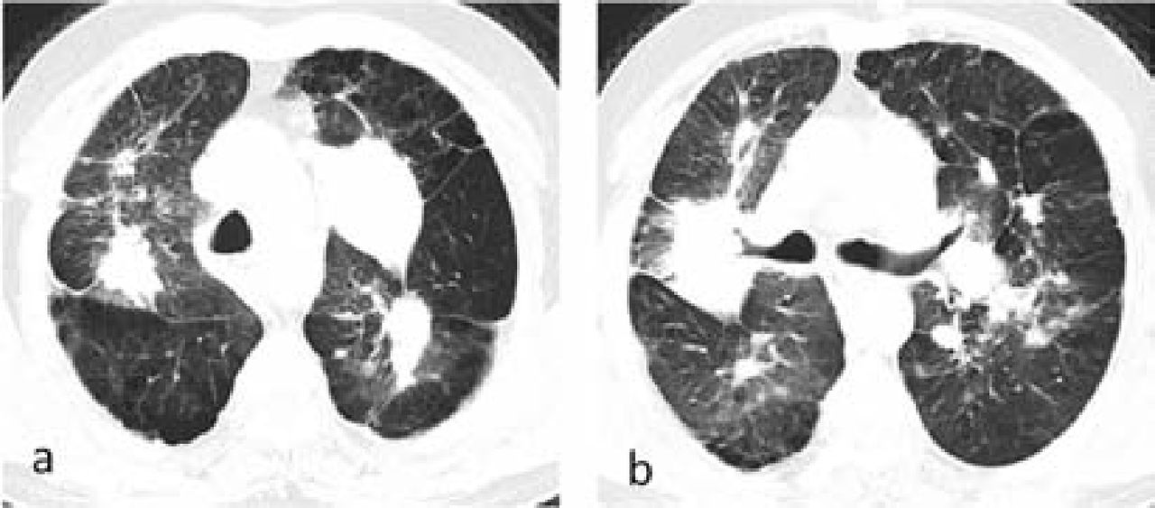
Axial CT (lung window) at the level of arch of (a) aorta and (b) carina reveals large bilateral masses with irregular margins indicative of PMF, as well as few small nodules and septal thickening. Also note the peripheral emphysematous changes. PMF = progressive massive fibrosis.
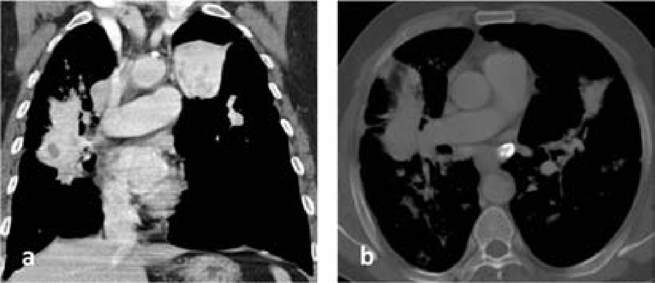
(a) Coronal CT obtained with mediastinal window settings reveals the masses to be of high density with few circumscribed low density areas; (b) Axial CT with bone window settings at the level of right pulmonary artery shows peripheral egg shell calcification in a subcarinal lymph node.
The occupational history and imaging findings were consistent with a diagnosis of complicated silicosis–progressive massive fibrosis (PMF). Silicosis is an occupational disorder caused by inhalation of microscopic silica dust particles in industries like mining and quarrying.1 On imaging, simple silicosis reveals multiple well defined small to medium-sized (<10 mm in diameter) centrilobular nodules in the upper lobe with enlarged mediastinal lymph nodes. Over time, the nodules migrate centrally and coalesce to form bilateral fibrotic masses which give an ‘angel-wing’ appearance on radiographs representing progressive massive fibrosis.2 Both the masses and nodes may show calcification.
There is no cure for PMF and treatment is mainly supportive. Lung transplantation may be considered. Serial chest radiographs should be obtained in patients with simple silicosis to monitor the development of PMF.
- © Royal College of Physicians 2015. All rights reserved.


