
Main menu

  • Home
  • Our journals
    • Clinical Medicine
    • Future Healthcare Journal
  • Subject collections
  • About the RCP
  • Contact us

Clinical Medicine Journal

  • ClinMed Home
  • Content
    • Current
    • Ahead of print
    • Archive
  • Author guidance
    • Instructions for authors
    • Submit online
  • About ClinMed
    • Scope
    • Editorial board
    • Policies
    • Information for reviewers
    • Advertising

User menu

  • Log in

Search

  • Advanced search
RCP Journals
Home
  • Log in
  • Home
  • Our journals
    • Clinical Medicine
    • Future Healthcare Journal
  • Subject collections
  • About the RCP
  • Contact us
Advanced

Clinical Medicine Journal

clinmedicine Logo
  • ClinMed Home
  • Content
    • Current
    • Ahead of print
    • Archive
  • Author guidance
    • Instructions for authors
    • Submit online
  • About ClinMed
    • Scope
    • Editorial board
    • Policies
    • Information for reviewers
    • Advertising

Image of the month: Radiological findings in bronchiectasis

Uzma Kamdar, Dilip Oswal and Salim Meghjee
Download PDF
DOI: https://doi.org/10.7861/clinmedicine.16-4-396
Clin Med August 2016
Uzma Kamdar
ADepartment of Respiratory Medicine, Pinderfields Hospital, Wakefield, UK
Roles: respiratory specialist registrar
  • Find this author on Google Scholar
  • Find this author on PubMed
  • Search for this author on this site
  • For correspondence: uzmakamdar@hotmail.com
Dilip Oswal
BDepartment of Radiology, Pinderfields Hospital, Wakefield, UK
Roles: consultant radiologist
  • Find this author on Google Scholar
  • Find this author on PubMed
  • Search for this author on this site
Salim Meghjee
CDepartment of Respiratory Medicine, Pinderfields Hospital, Wakefield, UK
Roles: consultant in respiratory medicine
  • Find this author on Google Scholar
  • Find this author on PubMed
  • Search for this author on this site
  • Article
  • Figures & Data
  • Info & Metrics
Loading

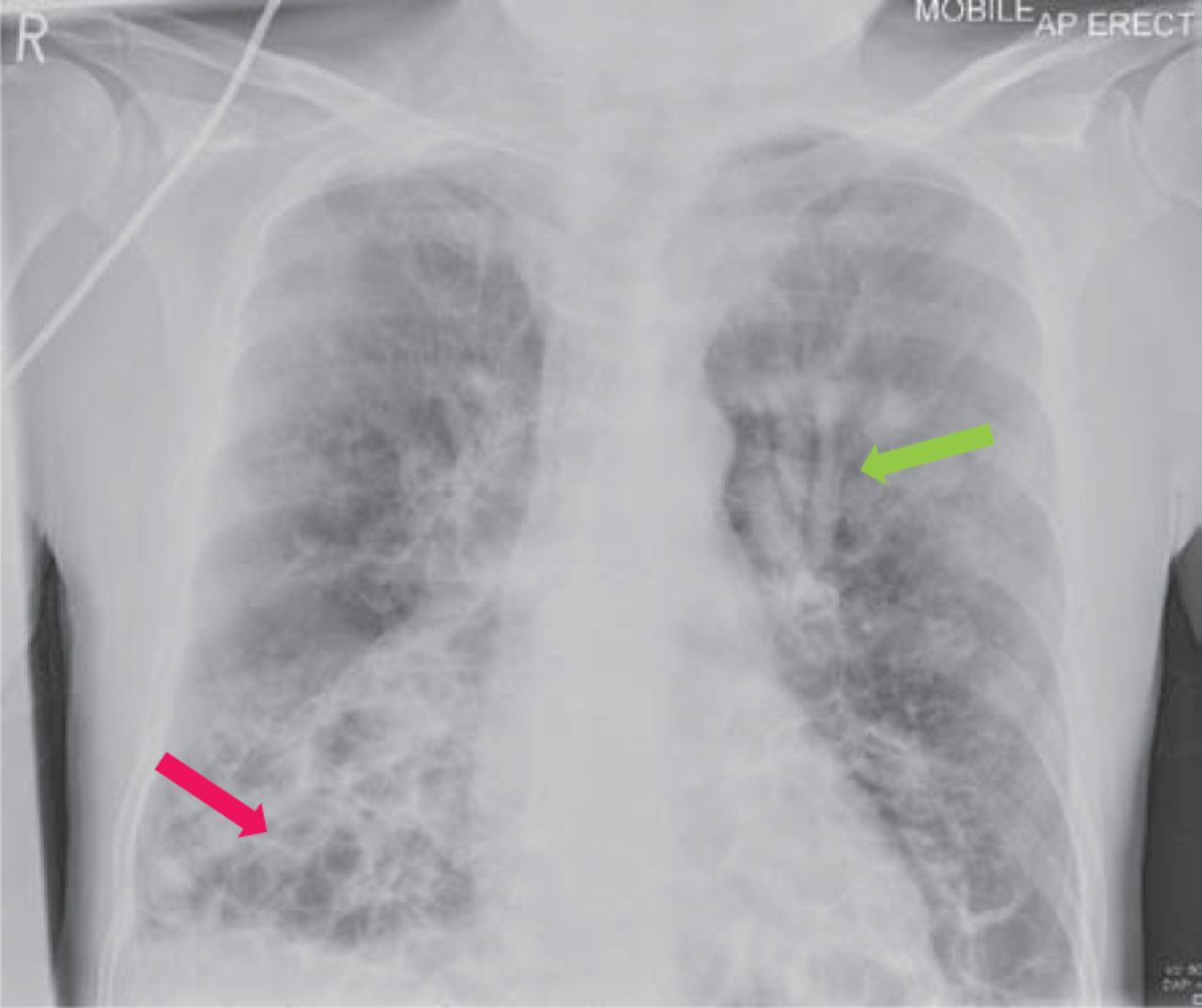
A 67-year-old patient with bronchiectasis, diagnosed at age 4, presented with worsening dyspnoea and hypoxia. Chest radiograph revealed finger in glove appearance in left lung and large cystic spaces at lung bases (Fig 1). A subsequent computed tomography scan ruled out pulmonary embolism; however, it showed the following interesting radiological features consistent with cystic-cylindrical bronchiectasis and bronchocoeles:

Fig 1.
  • Download figure
  • Open in new tab
  • Download powerpoint
Fig 1.

Chest X-ray showing finger in glove sign (green arrow) and cystic spaces at lung bases (red arrow).

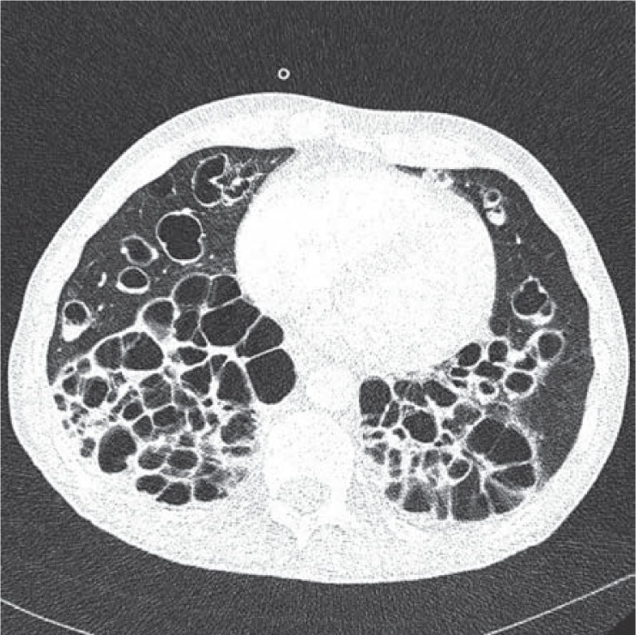
  1. Bunch of grapes sign1 – refers to dilated bronchi lying in close approximation giving the appearance of clusters of thin walled cysts (Figs 2 and 3). Similar appearances have also been described in hydatidiform mole and intraductal papillary mucinous neoplasm.

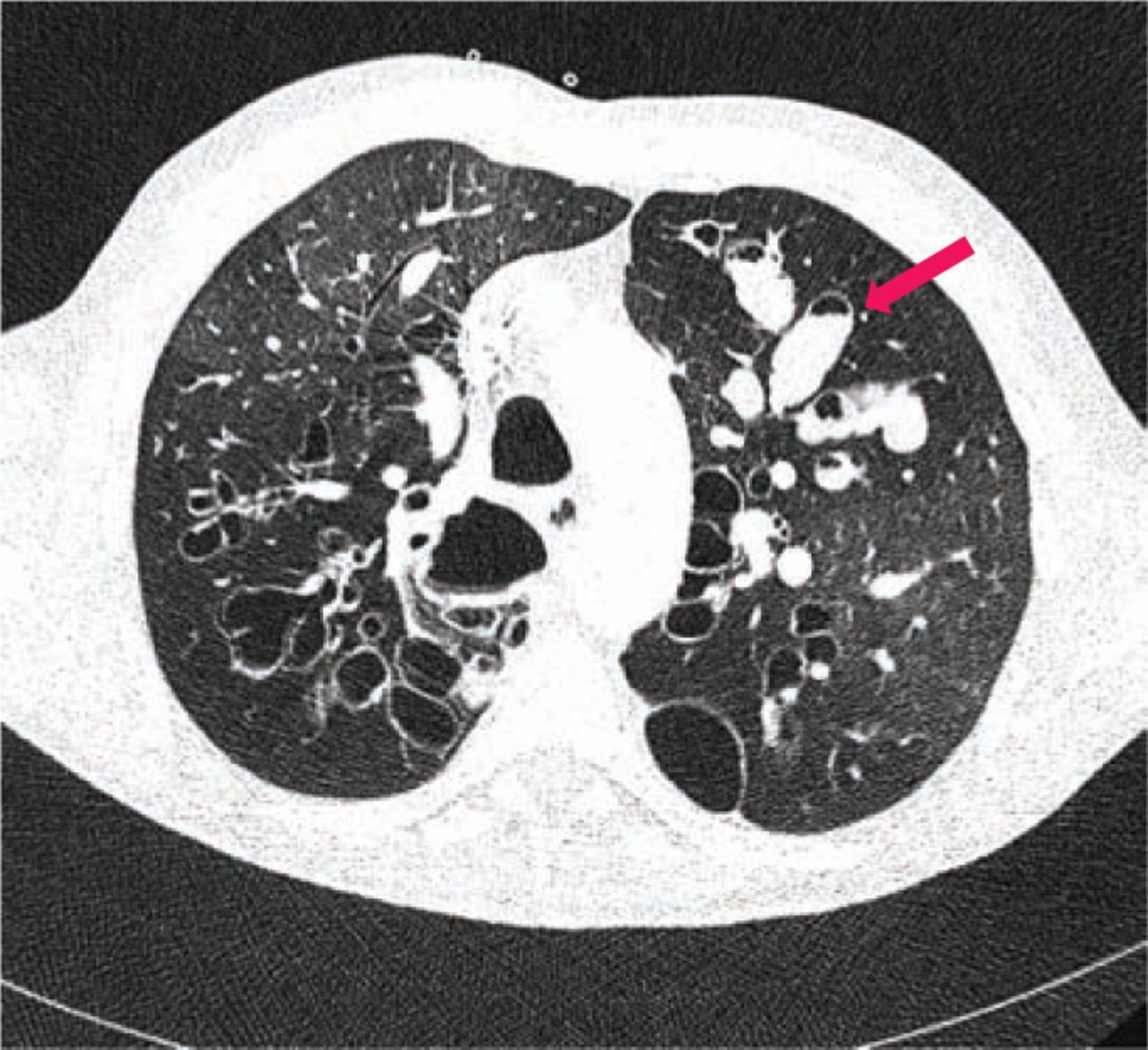
  2. Finger in glove sign2 – refers to dilated bronchi filled with mucus/mucoid impaction and are seen as opacities originating from hila directed peripherally (Figs 2 and 4).

  3. Bronchial arterial hypertrophy due to broncho-pulmonary shunting (Fig 5).

Fig 2.
  • Download figure
  • Open in new tab
  • Download powerpoint
Fig 2.

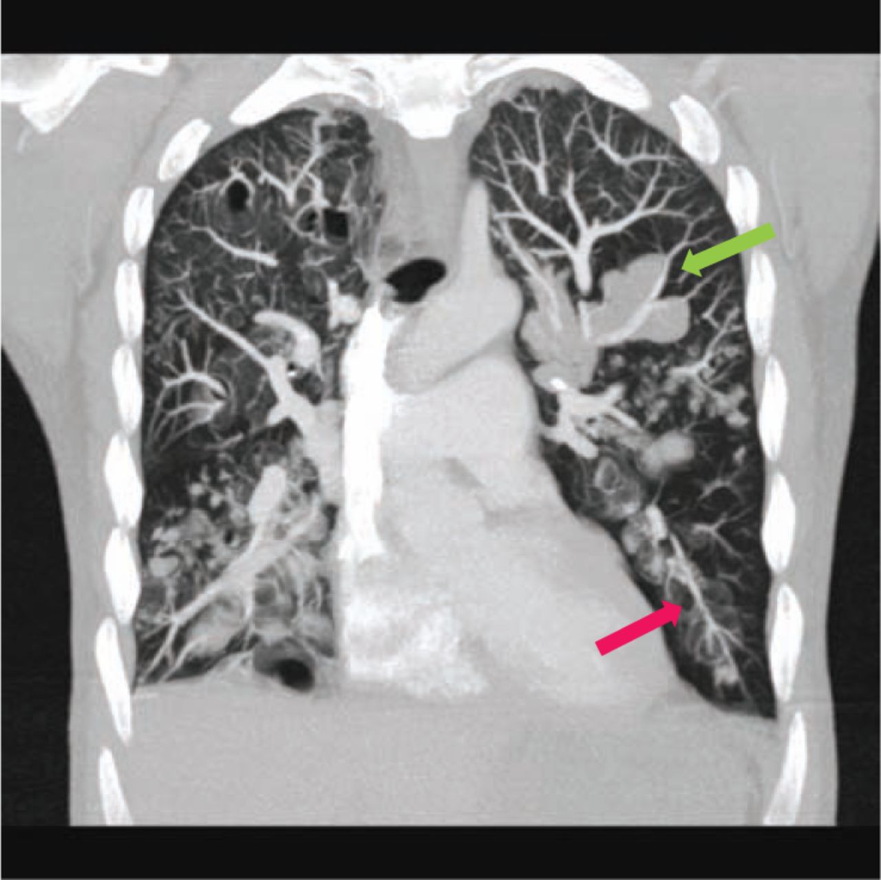
Coronal section of CT thorax showing finger in glove (green arrow) and bunch of grapes sign (red arrow).

Fig 3.
  • Download figure
  • Open in new tab
  • Download powerpoint
Fig 3.

CT section showing clusters of thin walled cystic spaces lying in close approximation ‘bunch of grape sign’.

Fig 4.
  • Download figure
  • Open in new tab
  • Download powerpoint
Fig 4.

CT section showing dilated bronchi filled with mucous (red arrow).

Fig 5.
  • Download figure
  • Open in new tab
  • Download powerpoint
Fig 5.

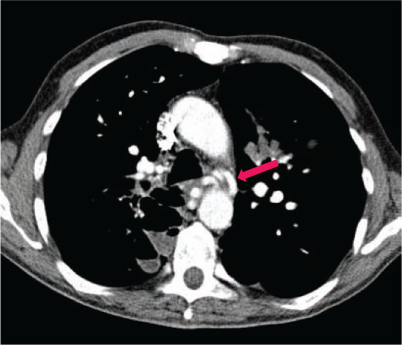
CT section showing bronchial artery hyperplasia (red arrow).

Competing interests

The authors declare no competing interests.

Acknowledgements

Written informed consent was obtained from the patient to publish the clinical details and images in this article.

  • © 2016 Royal College of Physicians

References

  1. ↵
    1. Naidich DP
    , Webb WR, Müller NL, Vlahos I, Krinsky GA. Computed tomography and magnetic resonance of the thorax, 4th edn. Philadelphia: Lippincott Williams & Wilkins, 2007.
  2. ↵
    1. Nguyen ET
    . The gloved finger sign. Radiology 2003;227:453–4.
    OpenUrlCrossRefPubMed
Back to top
Previous articleNext article

Article Tools

Download PDF
Article Alerts
Sign In to Email Alerts with your Email Address
Citation Tools
Image of the month: Radiological findings in bronchiectasis
Uzma Kamdar, Dilip Oswal, Salim Meghjee
Clinical Medicine Aug 2016, 16 (4) 396-397; DOI: 10.7861/clinmedicine.16-4-396

Citation Manager Formats

  • BibTeX
  • Bookends
  • EasyBib
  • EndNote (tagged)
  • EndNote 8 (xml)
  • Medlars
  • Mendeley
  • Papers
  • RefWorks Tagged
  • Ref Manager
  • RIS
  • Zotero
Share
Image of the month: Radiological findings in bronchiectasis
Uzma Kamdar, Dilip Oswal, Salim Meghjee
Clinical Medicine Aug 2016, 16 (4) 396-397; DOI: 10.7861/clinmedicine.16-4-396
del.icio.us logo Digg logo Reddit logo Twitter logo CiteULike logo Facebook logo Google logo Mendeley logo
  • Tweet Widget
  • Facebook Like
  • Google Plus One

Jump to section

  • Article
    • Competing interests
    • Acknowledgements
    • References
  • Figures & Data
  • Info & Metrics

Related Articles

  • No related articles found.
  • PubMed
  • Google Scholar

Cited By...

  • No citing articles found.
  • Google Scholar

More in this TOC Section

  • Images of the month: The conundrum of chronic coccidioidomycosis
  • Images of the month: Pulmonary cement embolism
  • Images of the month: Gliptin-induced bullous pemphigoid
Show more Image of the month

Similar Articles

Navigate this Journal

  • Journal Home
  • Current Issue
  • Ahead of Print
  • Archive

Related Links

  • ClinMed - Home
  • FHJ - Home
clinmedicine Footer Logo
  • Home
  • Journals
  • Contact us
  • Advertise
HighWire Press, Inc.

Follow Us:

  • Follow HighWire Origins on Twitter
  • Visit HighWire Origins on Facebook

Copyright © 2021 by the Royal College of Physicians