
Main menu

  • Home
  • Our journals
    • Clinical Medicine
    • Future Healthcare Journal
  • Subject collections
  • About the RCP
  • Contact us

Clinical Medicine Journal

  • ClinMed Home
  • Content
    • Current
    • Ahead of print
    • Archive
  • Author guidance
    • Instructions for authors
    • Submit online
  • About ClinMed
    • Scope
    • Editorial board
    • Policies
    • Information for reviewers
    • Advertising

User menu

  • Log in

Search

  • Advanced search
RCP Journals
Home
  • Log in
  • Home
  • Our journals
    • Clinical Medicine
    • Future Healthcare Journal
  • Subject collections
  • About the RCP
  • Contact us
Advanced

Clinical Medicine Journal

clinmedicine Logo
  • ClinMed Home
  • Content
    • Current
    • Ahead of print
    • Archive
  • Author guidance
    • Instructions for authors
    • Submit online
  • About ClinMed
    • Scope
    • Editorial board
    • Policies
    • Information for reviewers
    • Advertising

Image of the month: Gallstone ileus

Chia-Ming Chang, Chian-Ze Peng, Yen-Chia Chen and Chorng-Kuang How
Download PDF
DOI: https://doi.org/10.7861/clinmedicine.17-3-284
Clin Med June 2017
Chia-Ming Chang
ATaipei Veterans General Hospital, Taipei, Taiwan and National Yang-Ming University School of Medicine, Taipei, Taiwan
Roles: clinical lecturer
  • Find this author on Google Scholar
  • Find this author on PubMed
  • Search for this author on this site
  • For correspondence: cyberhunter700403@yahoo.com.tw
Chian-Ze Peng
BTaipei Veterans General Hospital, Taipei, Taiwan, Yuanshan & Su-Ao Branch, Yilan, Taiwan and National Yang-Ming University School of Medicine, Taipei, Taiwan
Roles: clinical lecturer
  • Find this author on Google Scholar
  • Find this author on PubMed
  • Search for this author on this site
Yen-Chia Chen
CTaipei Veterans General Hospital, Taipei, Taiwan and National Yang-Ming University School of Medicine, Taipei, Taiwan
Roles: assistant professor
  • Find this author on Google Scholar
  • Find this author on PubMed
  • Search for this author on this site
Chorng-Kuang How
DTaipei Veterans General Hospital, Taipei, Taiwan and National Yang-Ming University School of Medicine, Taipei, Taiwan
Roles: associate professor
  • Find this author on Google Scholar
  • Find this author on PubMed
  • Search for this author on this site
  • Article
  • Figures & Data
  • Info & Metrics
Loading

A 97-year-old woman presented to our emergency department with nausea, vomiting and abdominal pain for about 2 days. She had a history of ultrasound-guided percutaneous cholecystostomy following chronic cholecystitis with a huge gallstone 1 year earlier (Fig 1). Her physical examination showed hypoactive bowel sound and tympanic abdominal percussion. The abdominal radiograph showed no specific findings, except a distended stomach and dilated proximal small bowel loop. A follow-up abdominal computerised tomography (CT) scan demonstrated a gallstone measuring about 4.6 × 3.3 cm in the proximal jejunum, which caused proximal gas distension of the bowel, consistent with gallstone ileus (Fig 1). The patient underwent urgent enterolithotomy and she had an uneventful recovery.

Fig 1.
  • Download figure
  • Open in new tab
  • Download powerpoint
Fig 1.

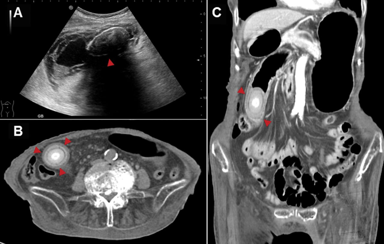
Gallstone image series. A – abdominal ultrasonography showing a huge gallstone within a thick-walled gallbladder (arrowhead); B and C – abdominal computerised tomography scan showing a calcified intra-luminal structure (arrowhead) in the proximal jejunum, which has caused proximal gas distension of the bowel, consistent with gallstone ileus.

Gallstone ileus is a rare bowel obstruction caused by cholelithiasis, which occurs more frequently in older women than men. The main cause of the development of a large gallstone in the intestine is a biliary enteric fistula. More than 70% of cases have cholecystoduodenal fistulas, followed by cholecystocolonic and cholecystogastric fistulas. Most gallstones affect the terminal ileum, which is the narrowest segment of the intestine.1

A preoperative diagnosis of gallstone ileus is challenging. Its nonspecific clinical presentation tends to delay diagnosis and treatment. The Rigler triad is the classic radiologic sign of gallstone ileus and consists of pneumobilia, intestinal obstruction and an ectopic gallstone. However, most gallstones are radiolucent and all three elements of the Rigler triad are present on plain radiographs in only 15% of cases. The Rigler triad is seen in up to 80% of cases when CT is used.2 Ultrasound is purported to have greater than 95% specificity and sensitivity in detecting gallstones within the gallbladder, but it is of limited use in cases of gallstone ileus because of gaseous distension of the bowel.3

Surgical intervention is the mainstay treatment strategy of gallstone ileus. The two-stage procedure (enterolithotomy followed by cholecystectomy and repair of the biliodigestive fistula after 4–6 weeks) is an alternative treatment suggested for patients with recurrent biliary symptoms.4 Nevertheless, a nonsurgical strategy, such as extracorporeal or electrohydraulic lithotripsy and the endoscopic removal of gallstones, has been reported.5n

Conflict of interest

The authors have no conflicts of interest to declare.

Acknowledgements

Written consent was obtained from the patient to publish the clinical details and images in this article.

  • © Royal College of Physicians 2017. All rights reserved.

References

  1. ↵
    1. Halabi WJ,
    2. Kang CY,
    3. Ketana N,
    4. et al.
    (2014) Surgery for gallstone ileus: a nationwide comparison of trends and outcomes. Ann Surg 259:329–35.
    OpenUrlCrossRefPubMed
  2. ↵
    1. Lassandro F,
    2. Romano S,
    3. Ragozzino A,
    4. et al.
    (2005) Role of helical CT in diagnosis of gallstone ileus and related conditions. AJR Am J Roentgenol 185:1159–65.
    OpenUrlCrossRefPubMed
  3. ↵
    1. Portincasa P,
    2. Moschetta A,
    3. Petruzzelli M,
    4. et al.
    (2006) Symptoms and diagnosis of gallbladder stones. Best Pract Res Clin Gastronenterol 20:1017–29.
    OpenUrl
  4. ↵
    1. Nuño-Guzmán CM,
    2. Marín-Contreras ME,
    3. Figueroa-Sánchez M,
    4. Corona JL.
    (2016) Gallstone ileus, clinical presentation, diagnostic and treatment approach. World J Gastronintest Surg 8:65–76.
    OpenUrl
  5. ↵
    1. Pezzoli A,
    2. Maimone A,
    3. Fusetti N,
    4. Pizzo E.
    (2015) Gallstone ileus treated with non-surgical conservative methods: a case report. J Med Case Rep 9:15.
    OpenUrl
Back to top
Previous articleNext article

Article Tools

Download PDF
Article Alerts
Sign In to Email Alerts with your Email Address
Citation Tools
Image of the month: Gallstone ileus
Chia-Ming Chang, Chian-Ze Peng, Yen-Chia Chen, Chorng-Kuang How
Clinical Medicine Jun 2017, 17 (3) 284-285; DOI: 10.7861/clinmedicine.17-3-284

Citation Manager Formats

  • BibTeX
  • Bookends
  • EasyBib
  • EndNote (tagged)
  • EndNote 8 (xml)
  • Medlars
  • Mendeley
  • Papers
  • RefWorks Tagged
  • Ref Manager
  • RIS
  • Zotero
Share
Image of the month: Gallstone ileus
Chia-Ming Chang, Chian-Ze Peng, Yen-Chia Chen, Chorng-Kuang How
Clinical Medicine Jun 2017, 17 (3) 284-285; DOI: 10.7861/clinmedicine.17-3-284
del.icio.us logo Digg logo Reddit logo Twitter logo CiteULike logo Facebook logo Google logo Mendeley logo
  • Tweet Widget
  • Facebook Like
  • Google Plus One

Jump to section

  • Article
    • Conflict of interest
    • Acknowledgements
    • References
  • Figures & Data
  • Info & Metrics

Related Articles

  • No related articles found.
  • PubMed
  • Google Scholar

Cited By...

  • No citing articles found.
  • Google Scholar

More in this TOC Section

  • Images of the month: The conundrum of chronic coccidioidomycosis
  • Images of the month: Pulmonary cement embolism
  • Images of the month: Gliptin-induced bullous pemphigoid
Show more Image of the month

Similar Articles

Navigate this Journal

  • Journal Home
  • Current Issue
  • Ahead of Print
  • Archive

Related Links

  • ClinMed - Home
  • FHJ - Home
clinmedicine Footer Logo
  • Home
  • Journals
  • Contact us
  • Advertise
HighWire Press, Inc.

Follow Us:

  • Follow HighWire Origins on Twitter
  • Visit HighWire Origins on Facebook

Copyright © 2021 by the Royal College of Physicians