
Article Figures & Data
Figures
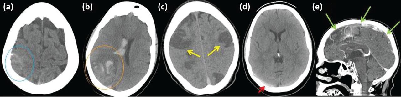
- Fig 1.
Non-contrast computerised tomography (CT) scans of brain. Demonstrating (a) hyperdensity over the right parietal cortex signifying subarachnoid haemorrhage (blue circle); (b) right occipital lobe haemorrhagic infarction with additional intraventricular haemorrhage (orange circle); (c) extensive and bilateral cerebral infarction (yellow arrows); and (d) hyperdensity in right transverse sinus demonstrating thrombosis (red arrow). (e) CT venogram demonstrating multiple areas of failed opacification of the superior sagittal sinus, representing thrombotic occlusion (green arrows)

Tables
- Table 1.
Signs and symptoms of cerebral venous thrombosis (CVT) and location of probable lesion
Signs and symptoms Probable lesion Headache Migraine Any venous occlusion/focal lesion Raised ICP a Large venous or sinus occlusion/large mass lesion Thunderclap Any venous occlusion/subarachnoid haemorrhage Ear/mastoid pain Transverse sinus with/without infection Focal neurological deficits Hemiparesis Infarction/haemorrhage/venous oedema Cranial nerve palsy III, IV Cavernous sinus V Cavernous sinus/superior petrosal sinus VI Cavernous sinus/inferior petrosal sinuses/raised ICP VII Transverse/sigmoid sinus VIII Transverse/sigmoid sinus/raised ICP IX, X, XI Posterior cavernous sinus/internal jugular vein/deep venous system Aphasia Focal infarction/haemorrhage/superficial or deep venous system Sensory disturbance Focal infarction/haemorrhage/superficial or deep venous system Inattention/neglect Focal infarction/haemorrhage/superficial venous system Ataxia Cerebellar veins/raised ICP Seizures Focal Focal infarction/haemorrhage Generalised Focal infarction/haemorrhage/severely raised ICP Visual disturbance Reduced acuity Raised ICP Reduced/altered visual field Raised ICP/Posterior infarction/haemorrhage/raised ICP (false localising sign) Diplopia Cavernous sinus/petrosal sinus/raised ICP Papilloedema Raised ICP Meningism Neck pain/stiffness Suggests infectious or inflammatory aetiology Photophobia Reduced consciousness Drowsiness Deep venous system/straight sinus/raised ICP/non-convulsive status epilepticus Stupor Coma Cognitive impairment Encephalopathy Deep venous system/temporal-parietal lesion (vein of Labbe)/seizures Disorientation Reduced concentration Amnesia ↵aRaised intracranial pressure (ICP) can result from a combination of a large venous/sinus occlusion or from large infarction/haemorrhage.
Thrombophilias Genetic – eg Factor V Leiden Acquired – eg antiphospholipid syndrome Infection Intracranial Regional eg ear, nose, throat, head, neck Systemic Trauma Head injury Cranial surgery Lumbar puncture Endovascular intervention Reproductive Pregnancy Puerperium Malignancy Intracranial Extracranial Medications Oral contraceptives Steroids Anti-neoplastic drugs (particularly L-asparaginase) Inflammatory Vasculitis eg Behçet’s disease Systemic lupus erythematosus Inflammatory bowel disease Sarcoidosis Haematological Iron deficiency anaemia Polycythaemia Endocrine Hyperthyroidism Systemic Dehydration Sepsis Intracranial abnormalities Dural fistulaeVenous anomalies Arteriovenous malformations


