
Main menu

  • Home
  • Our journals
    • Clinical Medicine
    • Future Healthcare Journal
  • Subject collections
  • About the RCP
  • Contact us

Clinical Medicine Journal

  • ClinMed Home
  • Content
    • Current
    • Ahead of print
    • Archive
  • Author guidance
    • Instructions for authors
    • Submit online
  • About ClinMed
    • Scope
    • Editorial board
    • Policies
    • Information for reviewers
    • Advertising

User menu

  • Log in

Search

  • Advanced search
RCP Journals
Home
  • Log in
  • Home
  • Our journals
    • Clinical Medicine
    • Future Healthcare Journal
  • Subject collections
  • About the RCP
  • Contact us
Advanced

Clinical Medicine Journal

clinmedicine Logo
  • ClinMed Home
  • Content
    • Current
    • Ahead of print
    • Archive
  • Author guidance
    • Instructions for authors
    • Submit online
  • About ClinMed
    • Scope
    • Editorial board
    • Policies
    • Information for reviewers
    • Advertising

Respiratory symptoms in a patient with inflammatory bowel disease

Jennifer Capps and Mark Woodhead
Download PDF
DOI: https://doi.org/10.7861/clinmedicine.18-2-186
Clin Med April 2018
Jennifer Capps
AWythenshawe Hospital, Manchester, UK
Roles: ST5 respiratory trainee
  • Find this author on Google Scholar
  • Find this author on PubMed
  • Search for this author on this site
  • For correspondence: Jennifer.capps@doctors.org.uk
Mark Woodhead
BManchester Royal Infirmary, Manchester, UK
Roles: respiratory consultant and honorary clinical professor of respiratory medicine
  • Find this author on Google Scholar
  • Find this author on PubMed
  • Search for this author on this site
  • Article
  • Figures & Data
  • Info & Metrics
Loading

A 47-year-old male presented with a 4-month history of breathlessness, chest discomfort and dry cough. He denied fevers, night sweats or weight loss. His medical history was of ulcerative colitis and hay fever. His current medications were azathioprine, mesalazine and infliximab (he had just received his third infusion). He had been screened and treated for latent tuberculosis (TB) prior to starting infliximab. He was a never smoker, an office worker, and kept no pets. He had no known family history, and was born in Pakistan. His most recent travel was 5 years ago.

The only abnormal findings on thoracic examination were bi-basal crackles. Routine blood tests were unremarkable. Three sputum samples were smear and culture negative for TB. A chest X-ray 1 year earlier was normal. A repeat chest X-ray was performed (Fig 1a).

Fig 1.
  • Download figure
  • Open in new tab
  • Download powerpoint
Fig 1.

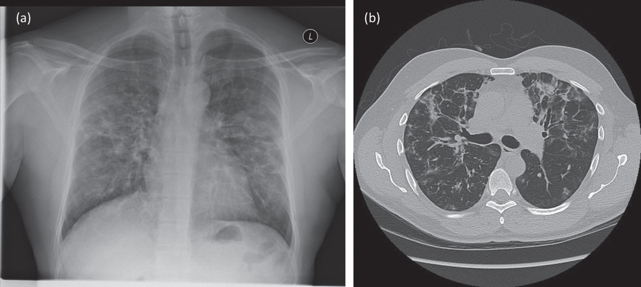
Baseline imaging (at presentation). (a) Chest X-ray. (b) CT thorax.

Possible diagnoses considered at this stage were infection (including TB), lung disease related to ulcerative colitis, and drug-induced interstitial lung disease.

A CT scan (Fig 1b) showed multilobar ground-glass consolidation with focal traction bronchiectasis, predominantly in upper and mid-zones, sparing the lung peripheries. There were also ground-glass foci within the bronchovascular bundles. There were small calcified lymph nodes in the mediastinum, but no lymphadenopathy elsewhere. A bronchoscopy with lavage was performed. Bronchoscopic appearances were normal and transbronchial biopsy from the lingula was normal. The lavage fluid was culture negative, and predominantly lymphocytic (61%).

A diagnosis of infliximab-related lung disease was made. Features supporting this include the lack of infective symptoms, the sterile sputum and the time course of the disease. In a large cohort of patients with rheumatoid arthritis, infliximab-related pneumonitis occurred after a mean of 2.8 infusions.1 The radiological appearances and lymphocytic bronchial lavage were in keeping with other reports of anti-TNF-related lung disease.2,3

The infliximab had been stopped shortly before respiratory review. Reports suggest the disease may resolve with cessation of the offending drug, but steroids are often given.2 In this case prednisolone 40 mg was initiated, and the patient’s symptoms resolved rapidly. Steroids were weaned over 5 months, and imaging following this (Fig 2a and 2b) showed resolution of most abnormalities, leaving scattered ground-glass foci and mild traction bronchiectasis.

Fig 2.
  • Download figure
  • Open in new tab
  • Download powerpoint
Fig 2.

Images following completion of steroids. (a) Chest X-ray. (b) CT thorax.

Consent

Patient consent was obtained to write and publish this article.

Acknowledgements

Thanks to Dr Devinda Karunaratne, consultant radiologist at Manchester Royal Infirmary, Manchester, who supplied the images included in this submission and provided a thoracic radiology opinion regarding the findings.

  • © Royal College of Physicians 2018. All rights reserved.

References

  1. ↵
    1. Takeuchi T
    , Tatsuki Y, Nogami Y, et al. Postmarketing surveillance of the safety profile of infliximab in 5000 Japanese patients with rheumatoid arthritis. Ann Rheum Dis 2008;67:189–94.
    OpenUrlAbstract/FREE Full Text
  2. ↵
    1. Lee H
    , Jo K, Shim TS, et al. Six cases of lung injury following antitumour necrosis factor therapy for inflammatory bowel disease. J Crohns Colitis 2015;9:1053–7.
    OpenUrlCrossRefPubMed
  3. ↵
    1. Weinstein EJ
    , Lykens MG. Pulmonary Complications Of Infliximab Therapy. Chest 2009;136:(4_MeetingAbstracts):35S.
    OpenUrl
Back to top
Previous articleNext article

Article Tools

Download PDF
Article Alerts
Sign In to Email Alerts with your Email Address
Citation Tools
Respiratory symptoms in a patient with inflammatory bowel disease
Jennifer Capps, Mark Woodhead
Clinical Medicine Apr 2018, 18 (2) 186-187; DOI: 10.7861/clinmedicine.18-2-186

Citation Manager Formats

  • BibTeX
  • Bookends
  • EasyBib
  • EndNote (tagged)
  • EndNote 8 (xml)
  • Medlars
  • Mendeley
  • Papers
  • RefWorks Tagged
  • Ref Manager
  • RIS
  • Zotero
Share
Respiratory symptoms in a patient with inflammatory bowel disease
Jennifer Capps, Mark Woodhead
Clinical Medicine Apr 2018, 18 (2) 186-187; DOI: 10.7861/clinmedicine.18-2-186
del.icio.us logo Digg logo Reddit logo Twitter logo CiteULike logo Facebook logo Google logo Mendeley logo
  • Tweet Widget
  • Facebook Like
  • Google Plus One

Jump to section

  • Article
    • Consent
    • Acknowledgements
    • References
  • Figures & Data
  • Info & Metrics

Related Articles

  • No related articles found.
  • PubMed
  • Google Scholar

Cited By...

  • No citing articles found.
  • Google Scholar

More in this TOC Section

  • Images of the month: The conundrum of chronic coccidioidomycosis
  • Images of the month: Pulmonary cement embolism
  • Images of the month: Gliptin-induced bullous pemphigoid
Show more Image of the month

Similar Articles

Navigate this Journal

  • Journal Home
  • Current Issue
  • Ahead of Print
  • Archive

Related Links

  • ClinMed - Home
  • FHJ - Home
clinmedicine Footer Logo
  • Home
  • Journals
  • Contact us
  • Advertise
HighWire Press, Inc.

Follow Us:

  • Follow HighWire Origins on Twitter
  • Visit HighWire Origins on Facebook

Copyright © 2021 by the Royal College of Physicians