
Main menu

  • Home
  • Our journals
    • Clinical Medicine
    • Future Healthcare Journal
  • Subject collections
  • About the RCP
  • Contact us

Clinical Medicine Journal

  • ClinMed Home
  • Content
    • Current
    • Ahead of print
    • Archive
  • Author guidance
    • Instructions for authors
    • Submit online
  • About ClinMed
    • Scope
    • Editorial board
    • Policies
    • Information for reviewers
    • Advertising

User menu

  • Log in

Search

  • Advanced search
RCP Journals
Home
  • Log in
  • Home
  • Our journals
    • Clinical Medicine
    • Future Healthcare Journal
  • Subject collections
  • About the RCP
  • Contact us
Advanced

Clinical Medicine Journal

clinmedicine Logo
  • ClinMed Home
  • Content
    • Current
    • Ahead of print
    • Archive
  • Author guidance
    • Instructions for authors
    • Submit online
  • About ClinMed
    • Scope
    • Editorial board
    • Policies
    • Information for reviewers
    • Advertising

Image of the month: Brachiocephalic arterial occlusive limb-shaking transient ischaemic attacks

Ahmed Abbas, Kurdow Nader, Alok Tiwari, Arunagiri Viruthagiri and Edward Littleton
Download PDF
DOI: https://doi.org/10.7861/clinmedicine.18-5-435
Clin Med October 2018
Ahmed Abbas
AQueen Elizabeth Hospital Birmingham, Birmingham, UK
Roles: ST6 clinical neurophysiology
  • Find this author on Google Scholar
  • Find this author on PubMed
  • Search for this author on this site
  • For correspondence: ahmed.abbas4@nhs.net
Kurdow Nader
BQueen Elizabeth Hospital Birmingham, Birmingham, UK
Roles: consultant neuroradiologist
  • Find this author on Google Scholar
  • Find this author on PubMed
  • Search for this author on this site
Alok Tiwari
CQueen Elizabeth Hospital Birmingham, Birmingham, UK
Roles: consultant vascular surgeon
  • Find this author on Google Scholar
  • Find this author on PubMed
  • Search for this author on this site
Arunagiri Viruthagiri
DQueen Elizabeth Hospital Birmingham, Birmingham, UK
Roles: senior vascular fellow
  • Find this author on Google Scholar
  • Find this author on PubMed
  • Search for this author on this site
Edward Littleton
EQueen Elizabeth Hospital Birmingham, Birmingham, UK
Roles: consultant neurologist
  • Find this author on Google Scholar
  • Find this author on PubMed
  • Search for this author on this site
  • Article
  • Figures & Data
  • Info & Metrics
Loading

A 75-year-old man with multiple cardiovascular risk factors presented with a 2-year history of increasingly frequent shaking episodes simultaneously affecting left upper and left lower limbs, lasting up to 30 seconds. There was no impairment of consciousness, speech or any other neurological disturbance in relation to these attacks but they were often precipitated by a change in posture. Limb-shaking transient ischaemic attacks (LS-TIA) were suspected.

Cervical doppler ultrasonography revealed significantly dampened flow in right common and internal carotid arteries. Computed tomography (CT) angiography showed dense calcification of the brachiocephalic (or innominate) artery, but no significant stenosis of the common or internal carotid arteries. Further assessment with invasive catheter angiography was undertaken, during which contrast injection into the aorta identified a proximally occluded brachiocephalic artery (Fig 1a). In addition, later downward filling of the distalmost brachiocephalic artery, and subsequently right subclavian artery, was illustrated by retrograde flow down the right cervical (common carotid and vertebral) arteries (Fig 1b). This retrograde flow, previously dubbed the ‘innominate steal phenomenon’,1 effectively reflected the right upper limb ‘stealing’ its arterial supply from the right cerebral hemisphere.

Fig 1.
  • Download figure
  • Open in new tab
  • Download powerpoint
Fig 1.

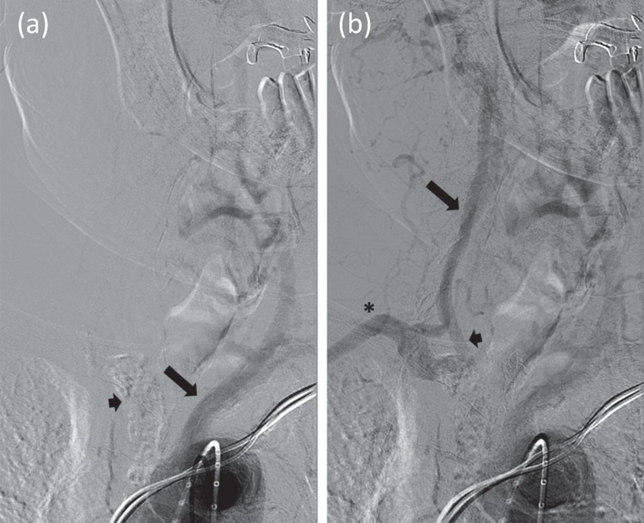
Catheter angiogram. (a) No filling of the calcified brachiocephalic artery (short arrow) despite filling of left common carotid artery (long arrow), followed shortly by (b) retrograde flow down the right vertebral artery (long arrow) and right common carotid artery (short arrow) to fill the distalmost brachiocephalic and ultimately right subclavian artery (asterisk).

An endovascular approach to revascularisation was deemed unsuitable due to heavy brachiocephalic calcification. Therefore, the patient underwent an extra-anatomical cervical left-to-right common carotid bypass procedure with a prosthetic graft. Postoperative recovery was uneventful and the procedure was successful in completely resolving his LS-TIA. Postoperative CT brain perfusion imaging also showed improved right hemispheric cerebral blood flow (Fig 2).

Fig 2.
  • Download figure
  • Open in new tab
  • Download powerpoint
Fig 2.

Computed tomography brain perfusion imaging. (a) Preoperative impaired right cerebral hemispheric blood flow (blue). (b) Postoperative improvement.

Almost all reports of LS-TIA relate to occlusive disease of the extracranial or intracranial carotid arterial system, with reduced forward arterial flow to the hemispheres.2,3 This case arising secondary to brachiocephalic arterial occlusion, with additional backward hemispheric arterial steal, is unusual and only one other case is recently reported in the literature.4 Awareness of extra-carotid arterial stenoses as potential causes of LS-TIA is important in accurately identifying the causally occluded arterial segment, and consequently in selecting the most appropriate surgical revascularisation procedure. In cases such as ours, wider diagnostic studies encompassing arterial vasculature proximal to the carotid arteries may be more sensitive in this regard compared to focal carotid arterial examinations.4

Consent

Consent was obtained from the patient to publish the clinical details and images in this article.

  • © Royal College of Physicians 2018. All rights reserved.

References

  1. ↵
    1. Uzun M
    , Bag˘cier S, Akkan K, et al. Innominate steal phenomenon: color and spectral Doppler sonographic findings. J Ultrasound Med 2008;27:1537–8.
    OpenUrlFREE Full Text
  2. ↵
    1. Baquis GD
    , Pessin MS, Scott RM. Limb shaking – a carotid TIA. Stroke 1985;16:444–8.
    OpenUrlAbstract/FREE Full Text
  3. ↵
    1. Salah Uddin AB
    . Limb shaking transient ischemic attack – an unusual presentation of carotid occlusive disease. A case report and review of the literature. Parkinsonism Relat Disord 2004;10:451–3.
    OpenUrlCrossRefPubMed
  4. ↵
    1. Goldstein ED
    , Cannistraro R, English S, et al. Brachiocephalic arterial occlusive disease presenting as limb-shaking transient ischemic attacks. J Stroke Cerebrovasc Dis 2018;27:e34–5.
    OpenUrl
Back to top
Previous articleNext article

Article Tools

Download PDF
Article Alerts
Sign In to Email Alerts with your Email Address
Citation Tools
Image of the month: Brachiocephalic arterial occlusive limb-shaking transient ischaemic attacks
Ahmed Abbas, Kurdow Nader, Alok Tiwari, Arunagiri Viruthagiri, Edward Littleton
Clinical Medicine Oct 2018, 18 (5) 435-436; DOI: 10.7861/clinmedicine.18-5-435

Citation Manager Formats

  • BibTeX
  • Bookends
  • EasyBib
  • EndNote (tagged)
  • EndNote 8 (xml)
  • Medlars
  • Mendeley
  • Papers
  • RefWorks Tagged
  • Ref Manager
  • RIS
  • Zotero
Share
Image of the month: Brachiocephalic arterial occlusive limb-shaking transient ischaemic attacks
Ahmed Abbas, Kurdow Nader, Alok Tiwari, Arunagiri Viruthagiri, Edward Littleton
Clinical Medicine Oct 2018, 18 (5) 435-436; DOI: 10.7861/clinmedicine.18-5-435
del.icio.us logo Digg logo Reddit logo Twitter logo Facebook logo Google logo Mendeley logo
  • Tweet Widget
  • Facebook Like
  • Google Plus One

Jump to section

  • Article
    • Consent
    • References
  • Figures & Data
  • Info & Metrics

Related Articles

  • No related articles found.
  • PubMed
  • Google Scholar

Cited By...

  • No citing articles found.
  • Google Scholar

More in this TOC Section

  • Images of the month 1: Ischaemic stroke due to pulmonary arteriovenous fistula
  • Images of the month 1: Histoacryl glue embolisation to the right ventricle following treatment for gastric varices
  • Images of the month 2: Disseminated nocardiosis in a 60-year-old woman with sarcoidosis
Show more Image of the month

Similar Articles

FAQs

  • Difficulty logging in.

There is currently no login required to access the journals. Please go to the home page and simply click on the edition that you wish to read. If you are still unable to access the content you require, please let us know through the 'Contact us' page.

  • Can't find the CME questionnaire.

The read-only self-assessment questionnaire (SAQ) can be found after the CME section in each edition of Clinical Medicine. RCP members and fellows (using their login details for the main RCP website) are able to access the full SAQ with answers and are awarded 2 CPD points upon successful (8/10) completion from:  https://cme.rcplondon.ac.uk

Navigate this Journal

  • Journal Home
  • Current Issue
  • Ahead of Print
  • Archive

Related Links

  • ClinMed - Home
  • FHJ - Home
clinmedicine Footer Logo
  • Home
  • Journals
  • Contact us
  • Advertise
HighWire Press, Inc.

Follow Us:

  • Follow HighWire Origins on Twitter
  • Visit HighWire Origins on Facebook

Copyright © 2021 by the Royal College of Physicians