
ABSTRACT
Synovitis, acne, pustulosis, hyperostosis and osteitis (SAPHO) syndrome is a rare, chronic, inflammatory disorder with cutaneous and osteoarticular manifestations.1 The aetiology of SAPHO syndrome is unknown and therefore treatment is tailored towards the individual. Non-steroidal anti-inflammatory drugs, bisphosphonates, corticosteriods, antibiotics, disease modifying anti-rheumatic drugs and biologics have all been used with variable success.
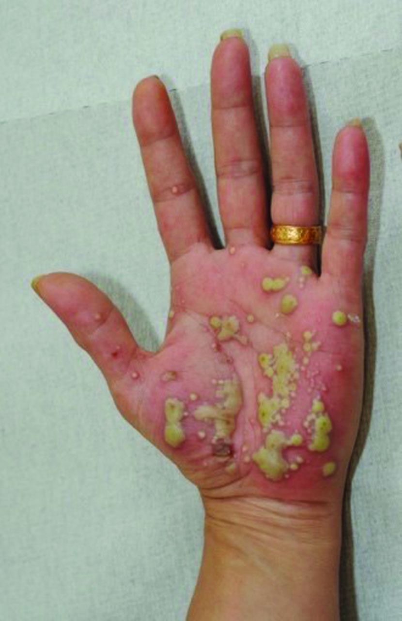
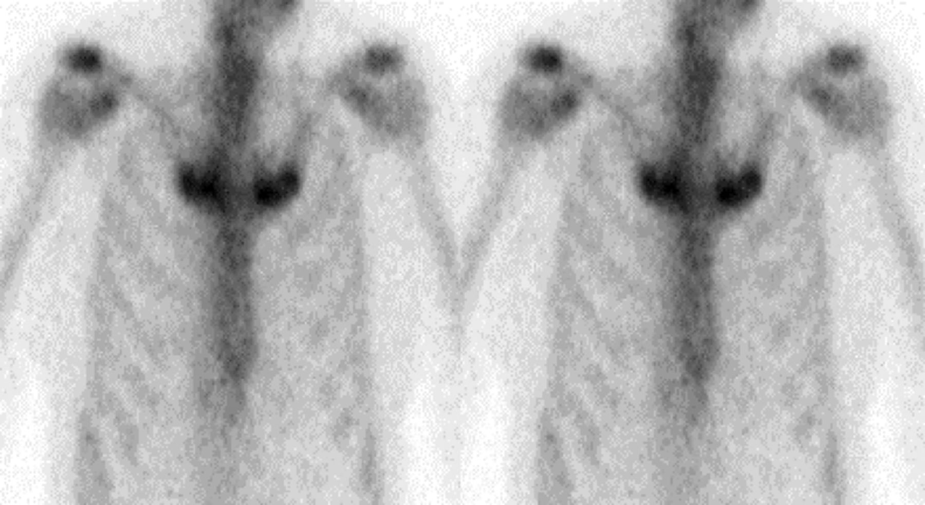
A 56-year-old woman presented with a 20-year history of palmoplantar pustulosis with anterior chest wall, shoulder and hip pain (Fig 1). Flare-ups of her symptoms appeared to coincide with stressful life events. Radiographs revealed sclerosis in the sacroiliac joints and bone scintigraphy revealed symmetric uptake in the sternoclavicular region with a ‘bull‘s head’ appearance pathognomonic of synovitis, acne, pustulosis, hyperostosis and osteitis (SAPHO) syndrome (Fig 2).1,2

Palmar pustulosis of the hand.

Bone scintigraphy showing intense uptake of the technetium-99m at the sternoclavicular joints and sternum representing the ‘bull's head’ sign.
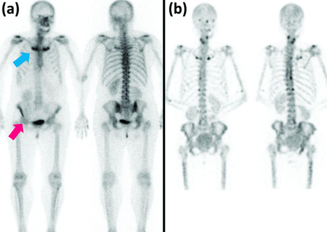
Initial treatment with ibuprofen and topical clobetasol proprionate 0.05% plus propylene glycol in a 60:40 ratio resulted in some symptomatic improvement. Subsequent trials with alendronic acid, methotrexate and ciclosporin were poorly tolerated and failed to alleviate her symptoms. Several cases have reported success with treating refractory SAPHO patients with anti-tumour necrosis factor-α (anti-TNF-α) agents, such as etanercept, infliximab and adalimumab. Although all have shown positive results in terms of improvement in articular symptoms, adalimumab has been shown to be superior in treating cutaneous symptoms.3 Consequently, our patient was commenced on adalimumab and after 1 year of treatment she has shown significant clinical and radiological improvements (Fig 3).

(a) Bone scintigraphy showing hyperfixation on sternoclavicular joints, both clavicles (blue arrow) and sacroiliac joints (red arrow) before starting treatment. (b) Bone scintigraphy showing significantly decreased uptake 4 years later after 1 year of treatment with adalimumab.
Radiological features are important in the diagnosis of SAPHO syndrome. While radiograms may demonstrate expanded bone, osteolysis, sclerosis, periosteal reaction and enthesopathic bone formation they often reveal no abnormalities at all.1 Bone scintigraphy is extremely valuable as increased uptake in affected bone determines if multiple sites are involved and helps to rule out malignancy and infection. Bone scintigraphy is also a useful way to monitor a patient's response to treatment and has emphasised the success of anti-TNF-α agents.
- © Royal College of Physicians 2019. All rights reserved.


Jump to section
Related Articles
Cited By...
- No citing articles found.