
Abstract
A 56-year-old woman presented with severe headache, blurring of vision, hypertensive emergency and severe crushing central chest pain. Extensive evaluation was undertaken to rule out sinister myocardial, pulmonary and mediastinal pathology. Pain relief required intravenous opiates. One week later, persistent complaints of numbness of hands and legs led to a suspicion of dysautonomic inflammatory neuropathy. Nerve conduction studies confirmed a demyelinating Guillain–Barré syndrome. Intravenous immunoglobulin treatment led to rapid resolution of pain, dysautonomia and neuropathic symptoms.
Case presentation
A 56-year-old woman presented to an outside hospital with sudden onset headache and blurring of vision. Her blood pressure (BP) was 220/120 mmHg. Magnetic resonance imaging (MRI) of the brain showed high parietal cortical diffusion-weighted (DW) MRI hyperintensities suggestive of a posterior reversible encephalopathy syndrome (PRES). She was admitted in the intensive care unit (ICU) and was treated with intravenous (IV) antihypertensives. On day 2, she developed episodes of severe crushing central chest pain, radiating to the back in approximately the T2–4 region lasting for up to an hour. She continued to have one to two more episodes of headache and blurring of vision. On day 3, she complained of numbness of her hands and soles. On day 4, she was transferred to our institution due to continuing episodes of chest pain, which occurred two to four times a day and required morphine or fentanyl infusion for control. At admission, a repeat MRI showed increase in the high cortical hyperintensities with new posterior occipital white matter hyperintensities compatible with PRES. On examination, she appeared mildly distressed, afebrile and her BP was 160/90 mmHg. Motor power was normal, she had diffuse areflexia and T3–4 vertebral tenderness.
Diagnosis
The initial differential diagnosis of ‘angina pectoris’ included myocardial or pericardial pathology, aortic dissection, pulmonary embolism and oesophageo-mediastinal lesions. Spinal cord pathology and radicular involvement were also high on the list (Box 1). Routine blood tests, HIV, venereal disease research laboratory and urine porphobilinogen were negative.
Differential diagnosis of angina pectoris
Investigations included serial electrocardiograms, echocardiogram, creatine kinase muscle/brain fraction serial troponins, transoesophageal echo (TEE), MRI whole spine with contrast, high-resolution computed tomography of the thorax and computed tomography aortogram. Upper gastrointestinal endoscopy showed mild oesophageal candidiasis and she was started on oral fluconazole. Barium swallow study showed normal barium follow through without any evidence of oesophageal spasm. She was started on intermittent morphine for the pain and double antiplatelets for possible angina. Her BP was controlled with IV labetalol and a continuous dexmedetomidine infusion was used for her agitation and pain.
On day 7, she complained of persistent numbness of her limbs and a nerve conduction study (NCS) was performed. This showed features of a mixed axonal and demyelinating neuropathy (Fig 1). At this point, Guillain–Barré syndrome (GBS) was considered.

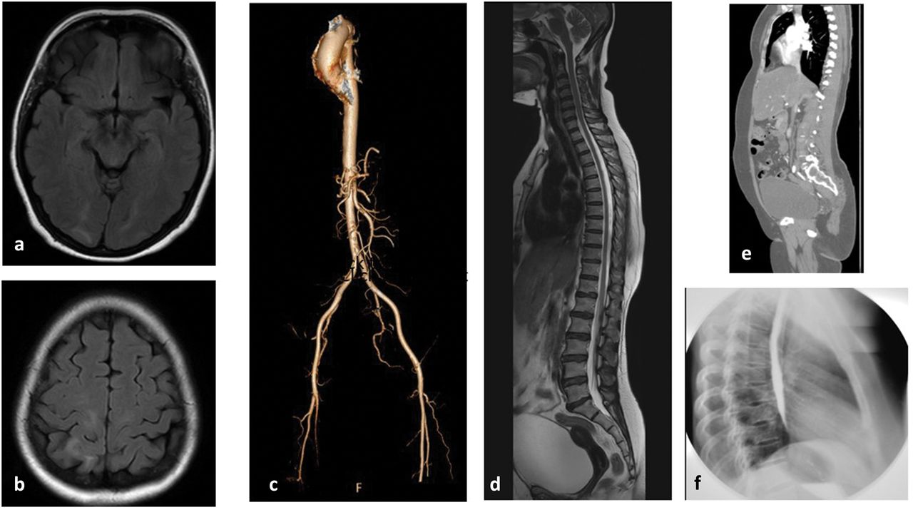
(a) and (b) Axial fluid-attenuated inversion recovery images showing posterior subcortical and gyral hyperintensities. (c) Computed tomography aortogram. (d) T2 sagittal whole spine magnetic resonance imaging. (e) Sagittal computed tomography thorax. (f) Normal barium swallow image (lateral).
Initial management and prognosis
She was started on IV immunoglobulin (Ig) 2 g/kg over 5 days. She required continuous dexmedetomidine for pain control as well as a labetalol infusion to control her BP and tachycardia for 7 days. Her numbness and pain started improving and she was ambulant by day 15 and transferred to the ward. Dysautonomia in GBS confers a guarded prognosis and clinicians must be attentive to the possibility of cardiac arrhythmias or sudden cardiac death.
Case progression and outcome
On day 15, a repeat NCS showed more delay in the distal motor latencies with an absent median, normal sural pattern compatible with a demyelinating neuropathy. On day 16, she was discharged, walking independently and with minimal residual glove and stocking numbness.
Discussion
GBS has rarely been associated with PRES syndrome. Only 14 cases have been reported in literature.1–3 PRES is a syndrome characterised by headache, altered sensorium, seizures, visual impairment and focal neurological deficits, and commonly associated with hypertension, eclampsia and a variety of conditions. MRI of the brain typically shows posterior (parieto-occipital white matter) symmetric vasogenic oedema that is hyperintense on T2-weighted image and fluid-attenuated inversion recovery. It is postulated to occur as a failure of cerebral autoregulatory mechanisms and secondary endothelial blood–brain barrier dysfunction. Most of the reported cases have been associated with dysautonomia (seen in ∼60% of GBS).4 Dysautonomia manifestations in GBS include tachy-bradycardia and fluctuating BP (Box 2). PRES can occur before, after or simultaneously with GBS manifestations. Interestingly, the vast majority of patients have been women aged >55 years (>90%) and most developed acute hypertension as in our case. PRES in GBS is associated with dysautonomia, high cerebrospinal fluid levels of inflammatory cytokines and chemokines or IV Ig administration. In most cases, supportive treatment of the BP has led to a good outcome in these cases.
Dysautonomic cardio- and cerebro-vascular complications of Guillain–Barré syndrome4
Chest pain in angina is mediated by myocardial tissue generation of adenosine during ischaemia. This activates sympathetic cardiac afferent neurones through the A1 subtype of the adenosine receptor.9 Unlike somatic pain, visceral pain (VP) is mediated by the autonomic nervous system (ANS) and is poorly localised. VP is carried by the afferent sympathetic nerves to the superior or inferior cardiac plexus and thence to the dorsal horn of the spinal cord. Here they ‘cross talk’ with somatic nociceptive fibres (from the upper cervical ganglion to T6–7 spinal cord segments). These also mix with afferent fibres from the tracheo-bronchial tree, lungs, oesophagus and stomach accounting for the extensive area of referred pain over the upper body in angina.10 Interestingly, our case is the first patient to present with severe crushing chest pain and PRES. The ‘anginal’ character led to a flurry of investigations to rule out sinister pathologies such as angina, mediastinitis and aortic dissection among others. Cardio-sympathetic hyperactivity in GBS is more common in the demyelinating type (acute inflammatory demyelinating polyneuropathy) type compared to the axonal variants such as acute motor axonal neuropathy or acute motor-sensory axonal neuropathy.11 Moreover, it can be disproportionate to the degree of sensori-motor involvement in GBS.
Summary
Neurogenic causes may present as an angina mimic.
Dysautonomia in GBS can cause both an ‘angina mimic’ and PRES.
Angina mimic can result from involvement of the thoracic sympathetic plexus.
Once the common causes of ‘angina’ are ruled out, autonomic testing or NCSs may be helpful to rule out a neuropathy.
- © 2019 Royal College of Physicians


Jump to section
Related Articles
Cited By...
- No citing articles found.