
Main menu

  • Home
  • Our journals
    • Clinical Medicine
    • Future Healthcare Journal
  • Subject collections
  • About the RCP
  • Contact us

Clinical Medicine Journal

  • ClinMed Home
  • Content
    • Current
    • Ahead of print
    • Archive
  • Author guidance
    • Instructions for authors
    • Submit online
  • About ClinMed
    • Scope
    • Editorial board
    • Policies
    • Information for reviewers
    • Advertising

User menu

  • Log in

Search

  • Advanced search
RCP Journals
Home
  • Log in
  • Home
  • Our journals
    • Clinical Medicine
    • Future Healthcare Journal
  • Subject collections
  • About the RCP
  • Contact us
Advanced

Clinical Medicine Journal

clinmedicine Logo
  • ClinMed Home
  • Content
    • Current
    • Ahead of print
    • Archive
  • Author guidance
    • Instructions for authors
    • Submit online
  • About ClinMed
    • Scope
    • Editorial board
    • Policies
    • Information for reviewers
    • Advertising

Images of the month: Lower-back pain in a young man with HLA B27 – not always spondyloarthritis!

Ashok Kumar, Ashish Sharma and Anunay Agarwal
Download PDF
DOI: https://doi.org/10.7861/clinmed.2020-0070
Clin Med May 2020
Ashok Kumar
AFortis Hospital, Noida, India
Roles: consultant
  • Find this author on Google Scholar
  • Find this author on PubMed
  • Search for this author on this site
  • For correspondence: ash.blueney@gmail.com
Ashish Sharma
BFortis Flt Lt Rajan Dhall Hospital, New Delhi, India
Roles: consultant and department head
  • Find this author on Google Scholar
  • Find this author on PubMed
  • Search for this author on this site
Anunay Agarwal
CAakash Hospital, New Delhi, India
Roles: consultant
  • Find this author on Google Scholar
  • Find this author on PubMed
  • Search for this author on this site
  • Article
  • Figures & Data
  • Info & Metrics
Loading

ABSTRACT

Presence of human leucocyte antigen (HLA) B27 in a patient with lower-back pain should not be considered synonymous with axial spondyloarthritis. Other causes of back pain should be ruled-out by careful evaluation. Lumbosacral transitional vertebra is a common congenital malformation of spine which frequently leads to mechanical back pain. We present a young man with lower-back pain who had HLA B27. He was labelled with axial spondyloarthritis elsewhere and was given etanercept without benefit. Further evaluation revealed that he had lumbosacral transitional vertebra and spina bifida occulta. No evidence of sacroiliitis was found. Etanercept was stopped and he was started on physiotherapy protocol for transitional vertebra, with which he improved remarkably. This case highlights the need for greater awareness among clinicians about lumbosacral transitional vertebra, a finding which is frequently missed. Presence of HLA B27 can be coincidental, as in our case.

KEYWORDS
  • Back pain
  • HLA B27
  • transitional vertebra
  • spina bifida occulta
  • spondyloarthritis

Case presentation

A 28-year-old man presented with lower-back pain, progressing over the previous 6 months. Forward bending and weight lifting aggravated the pain; rest provided relief. Human leucocyte antigen (HLA) B27 was positive and he was prescribed etanercept, subcutaneous injections of 50 mg weekly, elsewhere. He presented to our hospital after taking 10 weekly injections of etanercept with no benefit. Tenderness was observed in the right sacroiliac joint area.

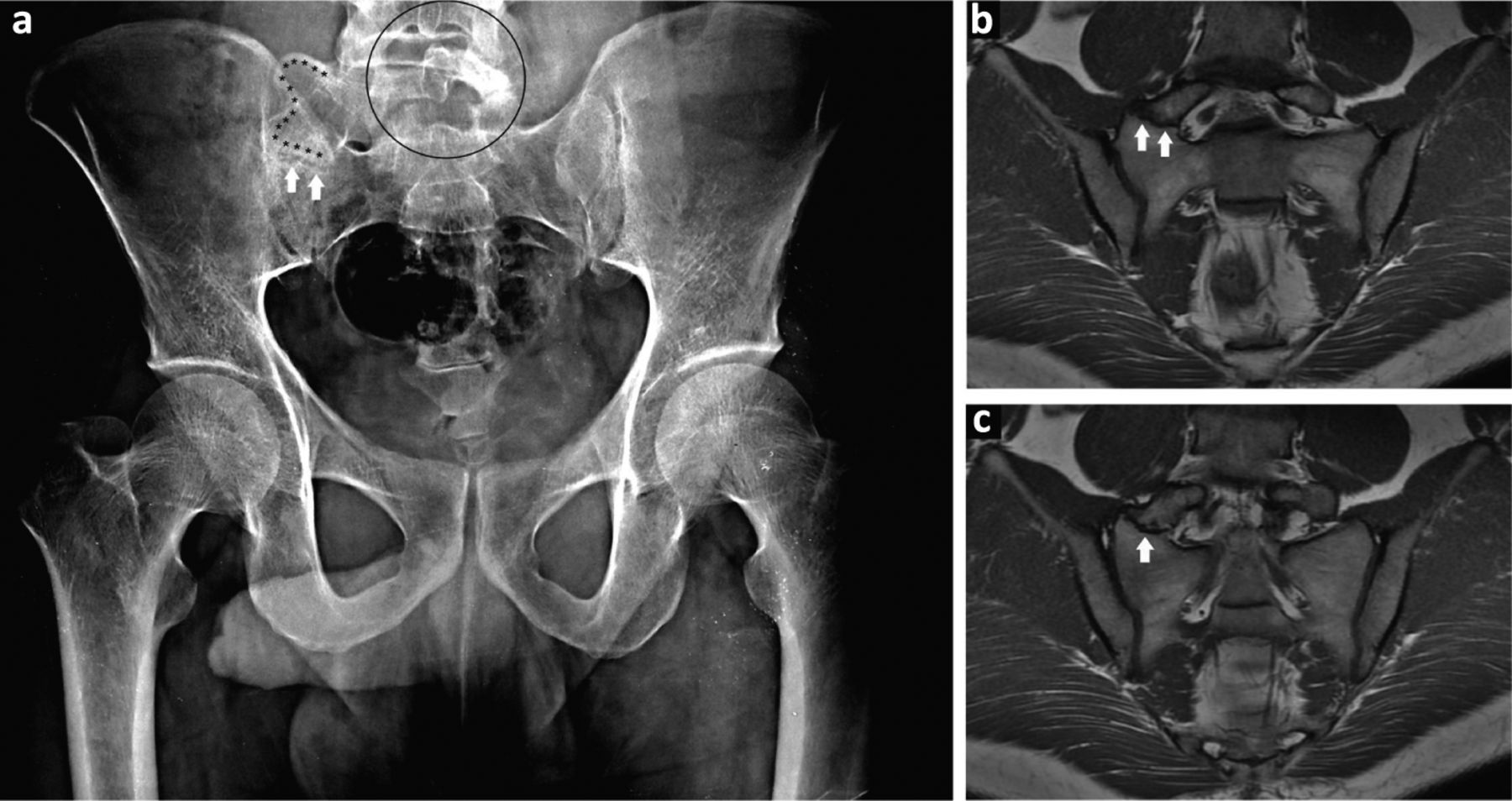
Investigations showed erythrocyte sedimentation rate of 12 mm in the first hour (normal range <30) and C-reactive protein of 2.8 mg per litre (normal range <10). X-ray of the pelvis revealed an elongated right transverse process of fifth lumbar vertebra and pseudoarthrosis with right ala of sacrum (Fig 1a). There was non-fusion of posterior elements of L5 vertebra, suggestive of spina bifida (Fig 1a). Magnetic resonance imaging (MRI) of pelvis confirmed the presence of pseudoarthrosis (Figs 1b and c); there was no sacroiliitis. The patient was diagnosed with lumbosacral transitional vertebra with spina bifida occulta. Etanercept was stopped and physiotherapy protocol for transitional vertebra was initiated. At the 4-month follow-up, the patient's back pain improved remarkably.

Fig 1.
  • Download figure
  • Open in new tab
  • Download powerpoint
Fig 1.

X-ray of pelvis and magnetic resonance imaging of sacroiliac joint showing lumbosacral transitional vertebra. a) X-ray of pelvis: anteroposterior projection showing elongated right transverse process of fifth lumbar vertebra (bordered by asterisks); pseudoarticulation between the right transverse process of L5 vertebra and ala of sacrum (arrows); non-fusion of posterior elements of L5 vertebra (encircled area) suggestive of spina bifida occulta. b) and c) Magnetic resonance imaging of sacroiliac joint: pseudoarticulation between the right transverse process of L5 vertebra and ala of sacrum (arrows).

Discussion

Lumbosacral transitional vertebra is a common congenital malformation of spine. Sacralisation of fifth lumbar vertebra or lumbarisation of first sacral vertebra is frequently observed. This results in pseudoarticulation between the ala of sacrum and the transverse process of the vertebra above.1 It leads to back pain due to intervertebral disc degeneration, facet joint arthropathy and mechanical stress at the level of pseudoarticulation.2 The pain may mimic that of inflammatory sacroiliitis due to axial spondyloarthritis. Our patient had HLA B27 in addition to lower-back pain, consequently, he was diagnosed with spondyloarthritis elsewhere. However, he did not have the inflammatory type of back pain and acute phase reactants and sacroiliac joints on MRI were normal. These points were against the diagnosis of spondyloarthritis. Non-steroidal anti-inflammatory drugs and physiotherapy are the mainstay of treatment.3

This case highlights the need for greater awareness among clinicians about lumbosacral transitional vertebra, a finding which is frequently missed. Presence of HLA B27 can be coincidental, as in our patient.

  • © Royal College of Physicians 2020. All rights reserved.

References

  1. ↵
    1. Jancuska JM
    , Spivak JM, Bendo JA. A review of symptomatic lumbosacral transitional vertebrae: Bertolotti's syndrome. Int J Spine Surg 2015;9:42.
    OpenUrlAbstract/FREE Full Text
  2. ↵
    1. Jain A
    , Agarwal A, Jain S, Shamshery C. Bertolotti syndrome: a diagnostic and management dilemma for pain physicians. Korean J Pain 2013;26:368–73.
    OpenUrl
  3. ↵
    1. Brenner AK.
    Use of lumbosacral region manipulation and therapeutic exercises for a patient with a lumbosacral transitional vertebra and low back pain. J Orthop Sports Phys Ther 2005;35:368–76.
    OpenUrlPubMed
Back to top
Previous articleNext article

Article Tools

Download PDF
Article Alerts
Sign In to Email Alerts with your Email Address
Citation Tools
Images of the month: Lower-back pain in a young man with HLA B27 – not always spondyloarthritis!
Ashok Kumar, Ashish Sharma, Anunay Agarwal
Clinical Medicine May 2020, 20 (3) e24-e25; DOI: 10.7861/clinmed.2020-0070

Citation Manager Formats

  • BibTeX
  • Bookends
  • EasyBib
  • EndNote (tagged)
  • EndNote 8 (xml)
  • Medlars
  • Mendeley
  • Papers
  • RefWorks Tagged
  • Ref Manager
  • RIS
  • Zotero
Share
Images of the month: Lower-back pain in a young man with HLA B27 – not always spondyloarthritis!
Ashok Kumar, Ashish Sharma, Anunay Agarwal
Clinical Medicine May 2020, 20 (3) e24-e25; DOI: 10.7861/clinmed.2020-0070
Reddit logo Twitter logo Facebook logo Mendeley logo
  • Tweet Widget
  • Facebook Like
  • Google Plus One

Jump to section

  • Article
    • ABSTRACT
    • Case presentation
    • Discussion
    • References
  • Figures & Data
  • Info & Metrics

Related Articles

  • No related articles found.
  • PubMed
  • Google Scholar

Cited By...

  • No citing articles found.
  • Google Scholar

More in this TOC Section

  • Images of the month 2: No laughing matter: Symptomatic Chiari malformation type I
  • Images of the month 1: Recurrent ischaemic stroke secondary to Eagle syndrome
  • Images of the month 2: Extensive pneumocephalus after subdural haematoma
Show more Images of the month

Similar Articles

FAQs

  • Difficulty logging in.

There is currently no login required to access the journals. Please go to the home page and simply click on the edition that you wish to read. If you are still unable to access the content you require, please let us know through the 'Contact us' page.

  • Can't find the CME questionnaire.

The read-only self-assessment questionnaire (SAQ) can be found after the CME section in each edition of Clinical Medicine. RCP members and fellows (using their login details for the main RCP website) are able to access the full SAQ with answers and are awarded 2 CPD points upon successful (8/10) completion from:  https://cme.rcplondon.ac.uk

Navigate this Journal

  • Journal Home
  • Current Issue
  • Ahead of Print
  • Archive

Related Links

  • ClinMed - Home
  • FHJ - Home
clinmedicine Footer Logo
  • Home
  • Journals
  • Contact us
  • Advertise
HighWire Press, Inc.

Follow Us:

  • Follow HighWire Origins on Twitter
  • Visit HighWire Origins on Facebook

Copyright © 2021 by the Royal College of Physicians