
Main menu

  • Home
  • Our journals
    • Clinical Medicine
    • Future Healthcare Journal
  • Subject collections
  • About the RCP
  • Contact us

Clinical Medicine Journal

  • ClinMed Home
  • Content
    • Current
    • Ahead of print
    • Archive
  • Author guidance
    • Instructions for authors
    • Submit online
  • About ClinMed
    • Scope
    • Editorial board
    • Policies
    • Information for reviewers
    • Advertising

User menu

  • Log in

Search

  • Advanced search
RCP Journals
Home
  • Log in
  • Home
  • Our journals
    • Clinical Medicine
    • Future Healthcare Journal
  • Subject collections
  • About the RCP
  • Contact us
Advanced

Clinical Medicine Journal

clinmedicine Logo
  • ClinMed Home
  • Content
    • Current
    • Ahead of print
    • Archive
  • Author guidance
    • Instructions for authors
    • Submit online
  • About ClinMed
    • Scope
    • Editorial board
    • Policies
    • Information for reviewers
    • Advertising

Images of the month: Verruciform xanthoma: an easily confused disease

Qian An, Li Zhang, Xing-Hua Gao and Song Zheng
Download PDF
DOI: https://doi.org/10.7861/clinmed.2019-0477
Clin Med July 2020
Qian An
AThe First Hospital of China Medical University, Shenyang, China
Roles: attending doctor
  • Find this author on Google Scholar
  • Find this author on PubMed
  • Search for this author on this site
Li Zhang
BThe First Hospital of China Medical University, Shenyang, China
Roles: associate professor
  • Find this author on Google Scholar
  • Find this author on PubMed
  • Search for this author on this site
Xing-Hua Gao
CThe First Hospital of China Medical University, Shenyang, China
Roles: professor
  • Find this author on Google Scholar
  • Find this author on PubMed
  • Search for this author on this site
Song Zheng
DThe First Hospital of China Medical University, Shenyang, China
Roles: associate professor of dermatology
  • Find this author on Google Scholar
  • Find this author on PubMed
  • Search for this author on this site
  • For correspondence: nyaadzs@163.com
  • Article
  • Figures & Data
  • Info & Metrics
Loading

ABSTRACT

Verruciform xanthomas occur primarily in the mouth, but sometimes in the anogenital region, which may cause clinical confusion with genital warts. We present a case where a 70-year-old man presented with an asymptomatic warty lesion on the scrotum and verruciform xanthoma was confirmed by histopathological results.

KEYWORDS
  • Verruciform
  • xanthoma

Case presentation

A 70-year-old man presented with an asymptomatic warty lesion on the scrotum, which initially developed as a small papule and enlarged into a polypoid nodule over 3 years. On examination, an exophytic yellowish verrucous nodule, measuring 3 cm in diameter, was found on the scrotum (Fig 1). The lesion was painless and moveable from the underlying structures. His past medical history was unremarkable and had no prior similar lesions on other sites.

Fig 1.
  • Download figure
  • Open in new tab
  • Download powerpoint
Fig 1.

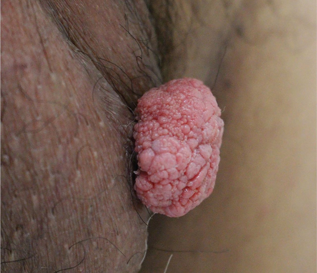
An exophytic yellowish verrucous nodule, measuring 3 cm in diameter, was found on the scrotum.

Investigations

Laboratory investigation for biochemical panel including serum lipid profile was within normal range. An excisional biopsy specimen revealed numerous foamy histiocytes in the papillary dermis (Fig 2a) which were positive for CD68 immunohistochemical staining (Fig 2b). Detection of human papillomavirus (HPV) deoxyribonucleic acid by polymerase chain reaction was negative.

Fig 2.
  • Download figure
  • Open in new tab
  • Download powerpoint
Fig 2.

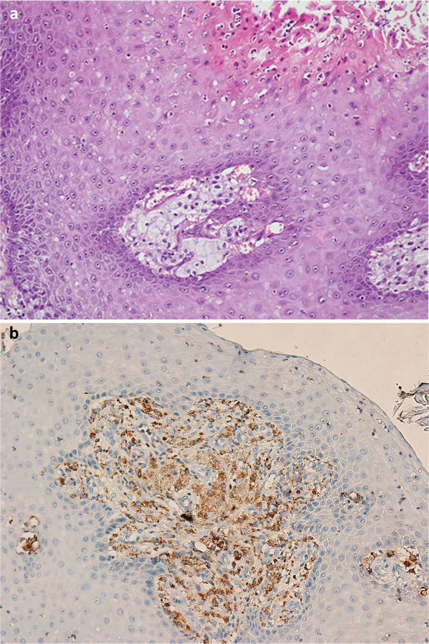
a) An excisional biopsy specimen revealed numerous foamy histiocytes in the papillary dermis. b) Foamy cells were positive for CD68 immunohistochemical staining.

Based on these findings, the diagnosis of verruciform xanthoma was made. The patient received a complete surgical excision of the lesion and had no recurrence at a 6-month follow-up.

Discussion

The exact aetiology and pathogenesis of verruciform xanthoma remains unknown. Several hypotheses have been proposed, including HPV infections or local release of lipids by damaged keratinocytes through chronic inflammation, though no HPV were detected in our patient.1 In contrast to other forms of xanthomas, there is usually no associated hyperlipidemia in verruciform xanthoma cases, as with our case.

Verruciform xanthomas occur primarily in the mouth, but sometimes in the anogenital (including the scrotum) region, which may cause clinical confusion with genital warts.2 Verruciform xanthomas should be differentiated with verrucous carcinoma, squamous cell carcinoma, bowenoid papulosis, granular cell tumor, giant molluscum contagiosum, verruca vulgaris and vulvar intraepithelial neoplasia. Therefore, the diagnosis of verruciform xanthomas should be confirmed by the histopathological results.

  • © Royal College of Physicians 2020. All rights reserved.

References

    1. Stiff KM
    , Cohen PR. Vegas (verruciform genital-associated) xanthoma: a comprehensive literature review. Dermatol Ther 2017;7: 65–79.
    OpenUrl
  1. ↵
    1. Joo J
    , Fung MA, Jagdeo J. Successful treatment of scrotal verruciform xanthoma with shave debulking and fractionated carbon dioxide laser therapy. Dermatol Surg 2014;40:214–7.
    OpenUrl
View Abstract
Back to top
Previous articleNext article

Article Tools

Download PDF
Article Alerts
Sign In to Email Alerts with your Email Address
Citation Tools
Images of the month: Verruciform xanthoma: an easily confused disease
Qian An, Li Zhang, Xing-Hua Gao, Song Zheng
Clinical Medicine Jul 2020, 20 (4) e123-e124; DOI: 10.7861/clinmed.2019-0477

Citation Manager Formats

  • BibTeX
  • Bookends
  • EasyBib
  • EndNote (tagged)
  • EndNote 8 (xml)
  • Medlars
  • Mendeley
  • Papers
  • RefWorks Tagged
  • Ref Manager
  • RIS
  • Zotero
Share
Images of the month: Verruciform xanthoma: an easily confused disease
Qian An, Li Zhang, Xing-Hua Gao, Song Zheng
Clinical Medicine Jul 2020, 20 (4) e123-e124; DOI: 10.7861/clinmed.2019-0477
del.icio.us logo Digg logo Reddit logo Twitter logo CiteULike logo Facebook logo Google logo Mendeley logo
  • Tweet Widget
  • Facebook Like
  • Google Plus One

Jump to section

  • Article
    • ABSTRACT
    • Case presentation
    • Investigations
    • Discussion
    • References
  • Figures & Data
  • Info & Metrics

Related Articles

  • No related articles found.
  • PubMed
  • Google Scholar

Cited By...

  • No citing articles found.
  • Google Scholar

More in this TOC Section

  • Image of the month: Placental transmogrification of the lung: a rare cause of cystic lung disease
  • Images of the month: An incidental finding of spontaneous pneumomediastinum (Hamman's syndrome) secondary to diabetic ketoacidosis during the coronavirus pandemic
  • Images of the month: Intrahepatic multiple low-signal lesions from Aeromonas salmonicida infection
Show more Images of the month

Similar Articles

Navigate this Journal

  • Journal Home
  • Current Issue
  • Ahead of Print
  • Archive

Related Links

  • ClinMed - Home
  • FHJ - Home
clinmedicine Footer Logo
  • Home
  • Journals
  • Contact us
  • Advertise
HighWire Press, Inc.

Follow Us:

  • Follow HighWire Origins on Twitter
  • Visit HighWire Origins on Facebook

Copyright © 2020 by the Royal College of Physicians