
Main menu

  • Home
  • Our journals
    • Clinical Medicine
    • Future Healthcare Journal
  • Subject collections
  • About the RCP
  • Contact us

Clinical Medicine Journal

  • ClinMed Home
  • Content
    • Current
    • Ahead of print
    • Archive
  • Author guidance
    • Instructions for authors
    • Submit online
  • About ClinMed
    • Scope
    • Editorial board
    • Policies
    • Information for reviewers
    • Advertising

User menu

  • Log in

Search

  • Advanced search
RCP Journals
Home
  • Log in
  • Home
  • Our journals
    • Clinical Medicine
    • Future Healthcare Journal
  • Subject collections
  • About the RCP
  • Contact us
Advanced

Clinical Medicine Journal

clinmedicine Logo
  • ClinMed Home
  • Content
    • Current
    • Ahead of print
    • Archive
  • Author guidance
    • Instructions for authors
    • Submit online
  • About ClinMed
    • Scope
    • Editorial board
    • Policies
    • Information for reviewers
    • Advertising

Images of the month: Crystalglobulin-induced nephropathy

Sam LF Lau, Fernand M-M Lai, Kai-Ming Chow and Cheuk-Chun Szeto
Download PDF
DOI: https://doi.org/10.7861/clinmed.2020-0116
Clin Med July 2020
Sam LF Lau
ACarol and Richard Yu Peritoneal Dialysis Research Centre, Hong Kong, China
Roles: fellow
  • Find this author on Google Scholar
  • Find this author on PubMed
  • Search for this author on this site
Fernand M-M Lai
BPrince of Wales Hospital, Hong Kong, China
Roles: professor
  • Find this author on Google Scholar
  • Find this author on PubMed
  • Search for this author on this site
Kai-Ming Chow
CCarol and Richard Yu Peritoneal Dialysis Research Centre, Hong Kong, China
Roles: consultant
  • Find this author on Google Scholar
  • Find this author on PubMed
  • Search for this author on this site
Cheuk-Chun Szeto
DCarol and Richard Yu Peritoneal Dialysis Research Centre, Hong Kong, China and The Chinese University of Hong Kong, Hong Kong, China
Roles: professor
  • Find this author on Google Scholar
  • Find this author on PubMed
  • Search for this author on this site
  • For correspondence: ccszeto@cuhk.edu.hk
  • Article
  • Figures & Data
  • Info & Metrics
Loading

ABSTRACT

We report a patient with chronic diabetes and was referred for recent onset proteinuria. Light microscopy of the renal biopsy specimen showed mildly expanded mesangium with mesangial hypercellularity and segmental sclerosis, features compatible with diabetic glomerulosclerosis. However, crystalglobulin-induced nephropathy with crystal deposit was identified on electron microscopy. Renal biopsy is often performed for diabetic patients who present with proteinuria and light microscopy often shows features of diabetic glomerulosclerosis. Additional information may occasionally be revealed on electron microscopy, altering the subsequent plan of management.

KEYWORDS
  • Glomerulonephritis
  • proteinuria

Case presentation

A 45-year-old man who had a history of hypertension, hyperlipidaemia and type 2 diabetes mellitus with retinopathy presented with frothy urine for 3 months. He had had diabetes for 4 years, and his medication included lisinopril 20 mg daily, amlodipine 10 mg daily, metformin 1,000 mg twice daily and glimepiride 1 mg every morning. Physical examination showed no oedema; his blood pressure was 140/80 mmHg, and the heart rate was 72 beats/min. Laboratory tests revealed a glycated haemoglobin (HbA1c) level of 7.1%, serum creatinine of 95 μmol/L, serum albumin of 33 g/L, total protein of 80 g/L and the spot urine total protein-to-creatinine ratio was 2.14 mg/mg-Cr. Further blood tests including hepatitis B surface antigen, anti-hepatitis C antibody, antinuclear antibody and anti-neutrophil cytoplasmic antibody were negative. Ultrasound showed normal-sized kidneys.

Investigation

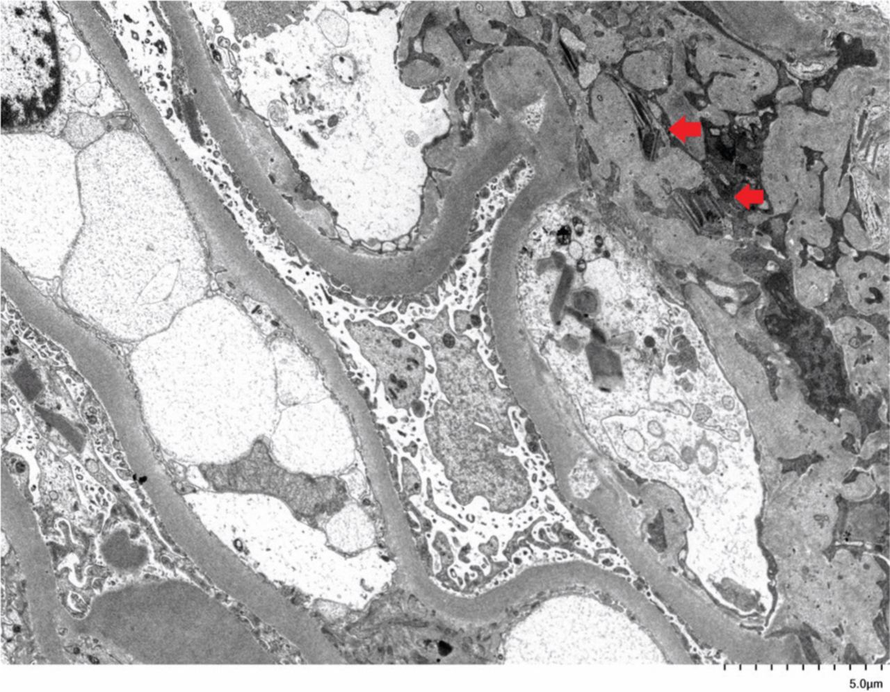
A renal biopsy was performed. Light microscopy of the renal biopsy specimen showed mildly expanded mesangium with mesangial hypercellularity and segmental sclerosis (Fig 1). There was no leukocyte infiltrate or necrotising lesion. Immunofluorescent study showed glomerular and tubular staining with predominantly kappa light chain. Electron microscopy revealed a uniformly thickened glomerular capillary basement membrane, prominent surface microvilli in podocytes with intracytoplasmic crystalline structure and intra-lysosomal inclusions (Fig 2). Similar crystalline cytoplasmic inclusion was also found in proximal tubules (not shown). Serum protein electrophoresis demonstrated immunoglobulin A (IgA) kappa paraproteinaemia 13 g/L, with a kappa-to-lambda ratio of 71 (reference range 0.26–1.65). Bone marrow examination showed 30–40% clonal plasmacytosis with kappa light chain restriction. There was no other clinical feature of Fanconi syndrome; serum uric acid, bicarbonate and phosphate levels were normal, although urine amino acid was not tested.

Fig 1.
  • Download figure
  • Open in new tab
  • Download powerpoint
Fig 1.

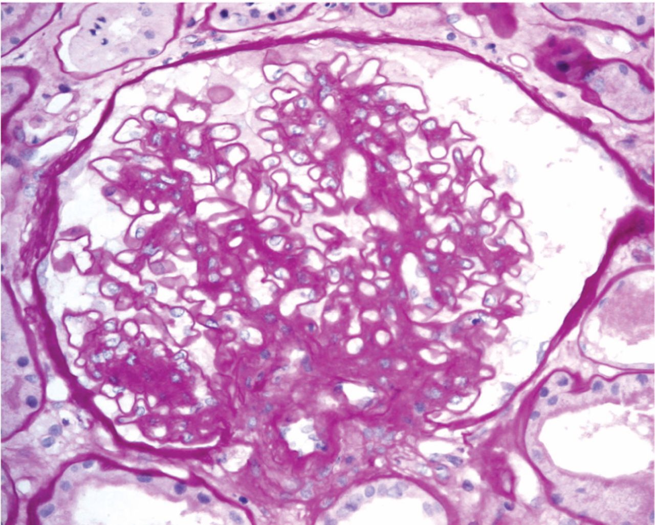
Light microscopy of renal biopsy with periodic acid–Schiff (PAS) stain showed mildly expanded mesangium with mesangial hypercellularity and segmental sclerosis.

Fig 2.
  • Download figure
  • Open in new tab
  • Download powerpoint
Fig 2.

Electron microscopy of renal biopsy showing crystalline structure (red arrows) in glomerular podocyte.

Discussion

Crystal nephropathies are classified according to the localisation of crystal deposits, namely the renal vasculature (type 1), the nephron (type 2) and the urinary tract (type 3).1 Crystal nephropathies may be caused by monoclonal gammopathy when the monoclonal immunoglobulin gives rise to the crystals.2 The clinical spectrum includes light chain proximal tubulopathy, crystal storing histiocytosis and crystalglobulin glomerulonephritis. Depending on the dynamics of crystal accumulation, either acute kidney injury or chronic kidney disease could occur.1

This case demonstrated the importance of electron microscopy in establishing the correct diagnosis. The IgA kappa paraproteinaemia and raised kappa to lambda ratio, which are often performed and available before renal biopsy, may give a clue to the underlying pathology. The identification of crystal deposit on electron microscopy is the key to the diagnosis. Bone marrow should be performed as the next step for clonal identification, in which plasma cells, lymphoid aggregates or amyloid deposits should be assessed. Imaging studies, such as computed tomography, should be considered especially when bone marrow examination is unrevealing and low-grade lymphoma is suspected. Clone-directed therapy is central to the management of monoclonal gammopathy of renal significance and bortezomib has the most robust data.3

  • © Royal College of Physicians 2020. All rights reserved.

References

  1. ↵
    1. Mulay SR
    , Anders HJ. Crystal nephropathies: mechanisms of crystal-induced kidney injury. Nat Rev Nephrol 2017;13:226–40.
    OpenUrlCrossRefPubMed
  2. ↵
    1. Leung N
    , Bridoux F, Batuman V, et al. The evaluation of monoclonal gammopathy of renal significance: a consensus report of the International Kidney and Monoclonal Gammopathy Research Group. Nat Rev Nephrol 2019;15:45–59.
    OpenUrl
  3. ↵
    1. Leung N
    , Drosou ME, Nasr SH. Dysproteinemias and Glomerular Disease. Clin J Am Soc Nephrol 2018;13:128–39.
    OpenUrlAbstract/FREE Full Text
Back to top
Previous articleNext article

Article Tools

Download PDF
Article Alerts
Sign In to Email Alerts with your Email Address
Citation Tools
Images of the month: Crystalglobulin-induced nephropathy
Sam LF Lau, Fernand M-M Lai, Kai-Ming Chow, Cheuk-Chun Szeto
Clinical Medicine Jul 2020, 20 (4) e125-e126; DOI: 10.7861/clinmed.2020-0116

Citation Manager Formats

  • BibTeX
  • Bookends
  • EasyBib
  • EndNote (tagged)
  • EndNote 8 (xml)
  • Medlars
  • Mendeley
  • Papers
  • RefWorks Tagged
  • Ref Manager
  • RIS
  • Zotero
Share
Images of the month: Crystalglobulin-induced nephropathy
Sam LF Lau, Fernand M-M Lai, Kai-Ming Chow, Cheuk-Chun Szeto
Clinical Medicine Jul 2020, 20 (4) e125-e126; DOI: 10.7861/clinmed.2020-0116
Reddit logo Twitter logo Facebook logo Mendeley logo
  • Tweet Widget
  • Facebook Like
  • Google Plus One

Jump to section

  • Article
    • ABSTRACT
    • Case presentation
    • Investigation
    • Discussion
    • References
  • Figures & Data
  • Info & Metrics

Related Articles

  • No related articles found.
  • PubMed
  • Google Scholar

Cited By...

  • No citing articles found.
  • Google Scholar

More in this TOC Section

  • Images of the month 2: No laughing matter: Symptomatic Chiari malformation type I
  • Images of the month 1: Recurrent ischaemic stroke secondary to Eagle syndrome
  • Images of the month 2: Extensive pneumocephalus after subdural haematoma
Show more Images of the month

Similar Articles

FAQs

  • Difficulty logging in.

There is currently no login required to access the journals. Please go to the home page and simply click on the edition that you wish to read. If you are still unable to access the content you require, please let us know through the 'Contact us' page.

  • Can't find the CME questionnaire.

The read-only self-assessment questionnaire (SAQ) can be found after the CME section in each edition of Clinical Medicine. RCP members and fellows (using their login details for the main RCP website) are able to access the full SAQ with answers and are awarded 2 CPD points upon successful (8/10) completion from:  https://cme.rcplondon.ac.uk

Navigate this Journal

  • Journal Home
  • Current Issue
  • Ahead of Print
  • Archive

Related Links

  • ClinMed - Home
  • FHJ - Home
clinmedicine Footer Logo
  • Home
  • Journals
  • Contact us
  • Advertise
HighWire Press, Inc.

Follow Us:

  • Follow HighWire Origins on Twitter
  • Visit HighWire Origins on Facebook

Copyright © 2021 by the Royal College of Physicians