
Main menu

  • Home
  • Our journals
    • Clinical Medicine
    • Future Healthcare Journal
  • Subject collections
  • About the RCP
  • Contact us

Clinical Medicine Journal

  • ClinMed Home
  • Content
    • Current
    • Ahead of print
    • Archive
  • Author guidance
    • Instructions for authors
    • Submit online
  • About ClinMed
    • Scope
    • Editorial board
    • Policies
    • Information for reviewers
    • Advertising

User menu

  • Log in

Search

  • Advanced search
RCP Journals
Home
  • Log in
  • Home
  • Our journals
    • Clinical Medicine
    • Future Healthcare Journal
  • Subject collections
  • About the RCP
  • Contact us
Advanced

Clinical Medicine Journal

clinmedicine Logo
  • ClinMed Home
  • Content
    • Current
    • Ahead of print
    • Archive
  • Author guidance
    • Instructions for authors
    • Submit online
  • About ClinMed
    • Scope
    • Editorial board
    • Policies
    • Information for reviewers
    • Advertising

Mechanisms of exertional angina in patients with normal coronary arteries

Haseeb Rahman, Ozan Demir, Matthew Ryan, Hannah McConkey, Howard Ellis, Cian Scannell, Amedeo Chiribiri, Andrew Webb and Divaka Perera
Download PDF
DOI: https://doi.org/10.7861/clinmed.20-2-s44
Clin Med March 2020
Haseeb Rahman
ASt Thomas’ Hospital, London, UK
  • Find this author on Google Scholar
  • Find this author on PubMed
  • Search for this author on this site
Ozan Demir
ASt Thomas’ Hospital, London, UK
  • Find this author on Google Scholar
  • Find this author on PubMed
  • Search for this author on this site
Matthew Ryan
ASt Thomas’ Hospital, London, UK
  • Find this author on Google Scholar
  • Find this author on PubMed
  • Search for this author on this site
Hannah McConkey
ASt Thomas’ Hospital, London, UK
  • Find this author on Google Scholar
  • Find this author on PubMed
  • Search for this author on this site
Howard Ellis
ASt Thomas’ Hospital, London, UK
  • Find this author on Google Scholar
  • Find this author on PubMed
  • Search for this author on this site
Cian Scannell
ASt Thomas’ Hospital, London, UK
  • Find this author on Google Scholar
  • Find this author on PubMed
  • Search for this author on this site
Amedeo Chiribiri
ASt Thomas’ Hospital, London, UK
  • Find this author on Google Scholar
  • Find this author on PubMed
  • Search for this author on this site
Andrew Webb
ASt Thomas’ Hospital, London, UK
  • Find this author on Google Scholar
  • Find this author on PubMed
  • Search for this author on this site
Divaka Perera
ASt Thomas’ Hospital, London, UK
  • Find this author on Google Scholar
  • Find this author on PubMed
  • Search for this author on this site
  • Article
  • Figures & Data
  • Info & Metrics
Loading

Background

Forty per cent of patients undergoing angiography to investigate exertional chest pain have normal coronary arteries. While described for nearly half a century, this condition has remained a mechanistic enigma.1–2 Diminished coronary blood flow augmentation to a pharmacological vasodilator, or coronary microvascular dysfunction (MVD), portends a greater risk of major adverse cardiovascular events.3–4 However, patients report symptoms during physical exercise, and the response to pharmacological ‘stress’ and physical exercise differ in the healthy heart.5 Moreover, it is unclear whether MVD is confined to the coronary circulation or a generalised disorder in myocardial and systemic blood flow during stress.

Aims

To identify whether patients with MVD have abnormal coronary blood flow during physical exercise, generalised systemic endothelial dysfunction and abnormal global myocardial blood flow during stress.

Method

Patients with angina and unobstructed coronary arteries underwent simultaneous acquisition of intra-coronary pressure and flow during rest, supine bicycle exercise and pharmacological vasodilatation with adenosine (hyperaemia), in the catheter laboratory. Coronary flow reserve (CFR) was calculated as hyperaemic coronary blood flow (CBF) / resting CBF.6 Wave intensity analysis quantified the proportion of accelerating wave energy (perfusion efficiency), a measure of the dynamic interaction between the contracting myocardium and interweaving coronary vasculature during the cardiac cycle, or cardiac–coronary coupling.5 Forearm blood flow ratio (FBF) was assessed to acetylcholine and NG-monomethyl-L-arginine (L-NMMA) infusions, an endothelial-dependent dilator and nitric oxide synthase inhibitor.7 Global myocardial blood flow and subendocardial:subepicardial perfusion ratio (endo/epi) were quantified using 3-Tesla cardiac magnetic resonance imaging (MRI) during hyperaemia and rest; inducible ischaemia was defined as hyperaemic endo/epi<1.0.8 Myocardial perfusion reserve (MPR) was calculated as hyperaemic myocardial blood flow / resting myocardial blood flow.9 Patients were classified as having MVD if CFR<2.5 and controls if CFR≥2.5, with researchers blinded to the classification.10

Results

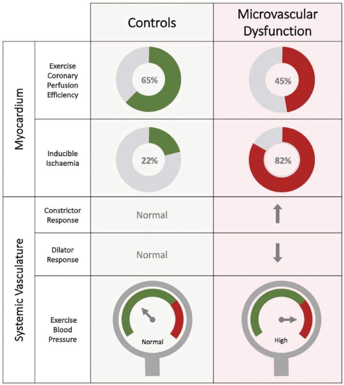
A total of 95 patients were enrolled (57±10 years, 81% women); 52 were classified as having MVD and 43 as controls. Microvascular resistance (MR) and CBF during peak exercise were similar in MVD and controls (4.5±1.6 vs 4.7±1.6 mmHg/cm/s and 30±10 vs 27±8 cm/s; p=0.68 and p=0.15). However, patients with MVD had higher systolic blood pressure and rate–pressure product (172±29 vs 155±29 mmHg and 20,486 vs 17,550 beats per minute*mmHg; both p=0.02). From rest to stress coronary perfusion efficiency improved in controls, whereas a paradoxical reduction occurred in those with MVD (59±11% to 65±14%; p=0.02 vs 61±12% to 44±10%; p<0.001; Fig 1). Patients with MVD had attenuated FBF augmentation to acetylcholine and exaggerated reduction to L-NMMA (3.6±1.8 vs 4.5±2.0 and 0.53±0.14 vs 0.78±0.09; p=0.02 and p<0.001). Those with MVD had a higher rate of inducible myocardial ischaemia and reduced MPR (22% vs 82% and 2.0±0.4 vs 2.7±0.5; both p<0.001).

Fig 1.
  • Download figure
  • Open in new tab
  • Download powerpoint
Fig 1.

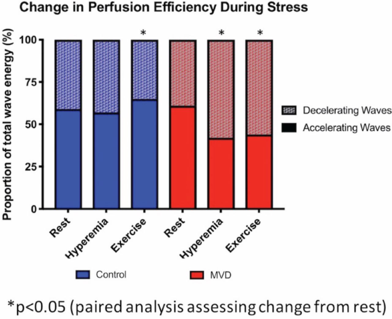
Changes in coronary perfusion efficiency (a measure of cardiac–coronary coupling) from rest to stress. MVD = microvascular dysfunction.

Conclusions

Compared with controls, MVD is associated with abnormal cardiac–coronary coupling during exercise, systemic endothelial dysfunction and hypertension with resultant increased myocardial oxygen demand. These pathophysiological changes act in concert to produce global myocardial ischaemia during stress and may underlie the higher incidence of cardiovascular morbidity observed in this population (Fig 2). The nitric oxide synthase pathway may represent a novel therapeutic target and should be the subject of future work; currently no disease-modifying therapy exists specifically for MVD.

Fig 2.
  • Download figure
  • Open in new tab
  • Download powerpoint
Fig 2.

Summary of cardiac and systemic responses to stress in microvascular dysfunction compared with controls.

Conflicts of interest

None declared.

  • © Royal College of Physicians 2020. All rights reserved.

References

  1. ↵
    1. Patel MR
    , Peterson ED, Dai D, et al. Low diagnostic yield of elective coronary angiography. N Engl J Med 2010;362:886–95.
    OpenUrlCrossRefPubMed
  2. ↵
    1. Likoff W
    , Segal BL, Kasparian H. Paradox of normal selective coronary arteriograms in patients considered to have unmistakable coronary heart disease. N Engl J Med 1967;276:1063–6.
    OpenUrlCrossRefPubMed
  3. ↵
    1. Pepine CJ
    , Anderson RD, Sharaf BL, et al. Coronary microvascular reactivity to adenosine predicts adverse outcome in women evaluated for suspected ischemia: results from the National Heart, Lung and Blood Institute. WISE (Women’s Ischemia Syndrome Evaluation) study. J Am Coll Cardiol 2010;55:2825–32.
    OpenUrlFREE Full Text
  4. ↵
    1. Taqueti VR
    , Shaw LJ, Cook NR, et al. Excess cardiovascular risk in women relative to men referred for coronary angiography is associated with severely impaired coronary flow reserve, not obstructive disease. Circulation 2017;135:566–77.
    OpenUrlAbstract/FREE Full Text
  5. ↵
    1. Lumley M
    , Williams R, Asrress KN, et al. Coronary physiology during exercise and vasodilation in the healthy heart and in severe aortic stenosis. J Am Coll Cardiol 2016;68:688–97.
    OpenUrlFREE Full Text
  6. ↵
    1. Gould KL
    , Lipscomb K, Hamilton GW. Physiologic basis for assessing critical coronary stenosis. Instantaneous flow response and regional distribution during coronary hyperemia as measures of coronary flow reserve. Am J Cardiol 1974;33:87–94.
    OpenUrlCrossRefPubMed
  7. ↵
    1. Khan SG
    , Geer A, Fok HW, et al. Impaired neuronal nitric oxide synthase-mediated vasodilator responses to mental stress in essential hypertension. Hypertension 2015;65:903–9.
    OpenUrl
  8. ↵
    1. Panting JR
    , Gatehouse PD, Yang GZ, et al. Abnormal subendocardial perfusion in cardiac syndrome X detected by cardiovascular magnetic resonance imaging. N Engl J Med 2002;346:1948–53.
    OpenUrlCrossRefPubMed
  9. ↵
    1. Schuster A
    , Sinclair M, Zarinabad N, et al. A quantitative high resolution voxel-wise assessment of myocardial blood flow from contrast-enhanced first-pass magnetic resonance perfusion imaging: microsphere validation in a magnetic resonance compatible free beating explanted pig heart model. Eur Heart J Cardiovasc Imaging 2015;16:1082–92.
    OpenUrlCrossRefPubMed
  10. ↵
    1. Ong P
    , Camici PG, Beltrame JF, et al. International standardization of diagnostic criteria for microvascular angina. Int J Cardiol 2018;250:16–20.
    OpenUrlPubMed
Back to top
Previous articleNext article

Article Tools

Download PDF
Article Alerts
Sign In to Email Alerts with your Email Address
Citation Tools
Mechanisms of exertional angina in patients with normal coronary arteries
Haseeb Rahman, Ozan Demir, Matthew Ryan, Hannah McConkey, Howard Ellis, Cian Scannell, Amedeo Chiribiri, Andrew Webb, Divaka Perera
Clinical Medicine Mar 2020, 20 (Suppl 2) s44-s45; DOI: 10.7861/clinmed.20-2-s44

Citation Manager Formats

  • BibTeX
  • Bookends
  • EasyBib
  • EndNote (tagged)
  • EndNote 8 (xml)
  • Medlars
  • Mendeley
  • Papers
  • RefWorks Tagged
  • Ref Manager
  • RIS
  • Zotero
Share
Mechanisms of exertional angina in patients with normal coronary arteries
Haseeb Rahman, Ozan Demir, Matthew Ryan, Hannah McConkey, Howard Ellis, Cian Scannell, Amedeo Chiribiri, Andrew Webb, Divaka Perera
Clinical Medicine Mar 2020, 20 (Suppl 2) s44-s45; DOI: 10.7861/clinmed.20-2-s44
del.icio.us logo Digg logo Reddit logo Twitter logo CiteULike logo Facebook logo Google logo Mendeley logo
  • Tweet Widget
  • Facebook Like
  • Google Plus One

Jump to section

  • Article
    • Background
    • Aims
    • Method
    • Results
    • Conclusions
    • Conflicts of interest
    • References
  • Figures & Data
  • Info & Metrics

Related Articles

  • No related articles found.
  • PubMed
  • Google Scholar

Cited By...

  • No citing articles found.
  • Google Scholar

More in this TOC Section

  • Adherence to therapy in chronic inflammatory rheumatic diseases: the older the better
  • A review in general practice: the appropriate prescription of PPIs with those taking aspirin over the age of 70
  • A rare case of ascites
Show more Clinical

Similar Articles

Navigate this Journal

  • Journal Home
  • Current Issue
  • Ahead of Print
  • Archive

Related Links

  • ClinMed - Home
  • FHJ - Home
clinmedicine Footer Logo
  • Home
  • Journals
  • Contact us
  • Advertise
HighWire Press, Inc.

Follow Us:

  • Follow HighWire Origins on Twitter
  • Visit HighWire Origins on Facebook

Copyright © 2021 by the Royal College of Physicians