
Main menu

  • Home
  • Our journals
    • Clinical Medicine
    • Future Healthcare Journal
  • Subject collections
  • About the RCP
  • Contact us

Clinical Medicine Journal

  • ClinMed Home
  • Content
    • Current
    • Ahead of print
    • Archive
  • Author guidance
    • Instructions for authors
    • Submit online
  • About ClinMed
    • Scope
    • Editorial board
    • Policies
    • Information for reviewers
    • Advertising

User menu

  • Log in

Search

  • Advanced search
RCP Journals
Home
  • Log in
  • Home
  • Our journals
    • Clinical Medicine
    • Future Healthcare Journal
  • Subject collections
  • About the RCP
  • Contact us
Advanced

Clinical Medicine Journal

clinmedicine Logo
  • ClinMed Home
  • Content
    • Current
    • Ahead of print
    • Archive
  • Author guidance
    • Instructions for authors
    • Submit online
  • About ClinMed
    • Scope
    • Editorial board
    • Policies
    • Information for reviewers
    • Advertising

Images of the month: Gliptin-induced bullous pemphigoid

Oluwafikunayo Orekoya and Ian H Coulson
Download PDF
DOI: https://doi.org/10.7861/clinmed.2020-0825
Clin Med January 2021
Oluwafikunayo Orekoya
ASalford Royal NHS Foundation Trust, Salford, UK
Roles: ST4 dermatology registrar
  • Find this author on Google Scholar
  • Find this author on PubMed
  • Search for this author on this site
Ian H Coulson
BBurnley General Hospital, Burnley, UK
Roles: consultant dermatologist
  • Find this author on Google Scholar
  • Find this author on PubMed
  • Search for this author on this site
  • Article
  • Figures & Data
  • Info & Metrics
Loading
KEYWORDS:
  • dermatology
  • bullous pemphigoid
  • gliptin,
  • DPP4-inhibitors

Case presentation

A 67-year-old woman with a background of type 2 diabetes mellitus (T2DM) presented to the dermatology department with a 4–6 week history of a blistering eruption and oral mucosal ulceration.

She had blisters arranged in an annular fashion and erosions on her trunk and limbs, and desquamative gingivitis and ulceration in her mouth (Fig 1). A diagnosis of bullous pemphigoid or linear immunoglobulin (Ig) A bullous dermatosis was suspected; the former was confirmed by histologic findings of a sub-epidermal blister with a polymorphonuclear infiltrate admixed with eosinophils and the presence of linear staining for IG3 and C3 at the basement membrane zone on direct immunofluorescence.

Fig 1.
  • Download figure
  • Open in new tab
  • Download powerpoint
Fig 1.

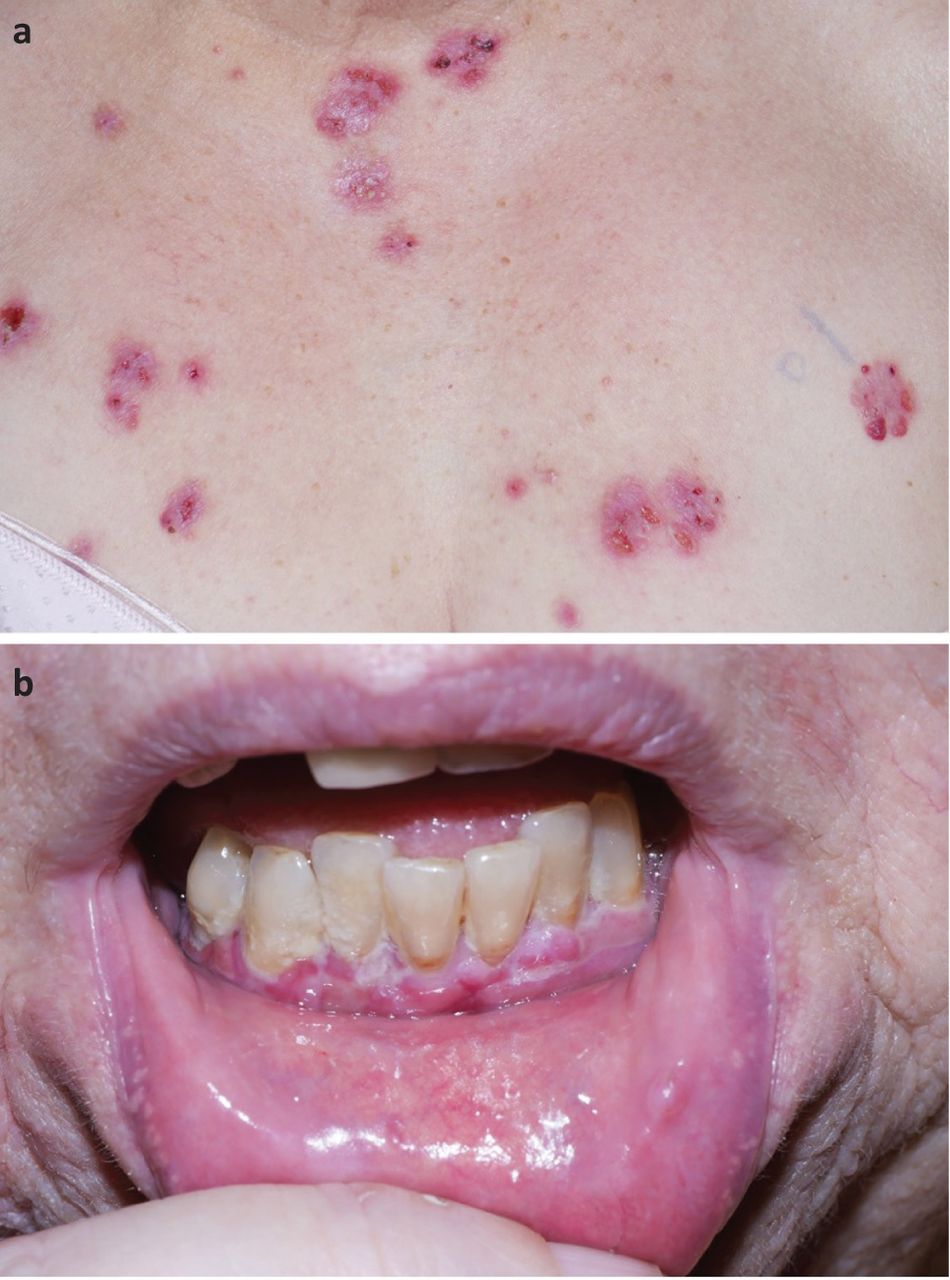
a) Blisters and erosions on the anterior chest wall. b) Desquamative gingivitis.

From her medication history, we identified that 13 months prior to developing the rash she was started on linagliptin, a dipeptidyl peptidase-4 (DPP-4) inhibitor, to manage her T2DM. The linagliptin was stopped and she was managed with topical corticosteroids, clobetasol propionate 0.05% ointment and betamethasone 500 mg mouthwash.

She improved without requiring systemic corticosteroids or other immunosuppressive agents. Interestingly, as a result of a painful mouth, she lost 15 kg in weight and no longer required oral anti-diabetic medication.

Discussion

Bullous pemphigoid is an organ specific autoimmune blistering skin disease that most commonly affects the elderly and has an annual incidence of 4.3 per 100,000.1 It occurs as a result of immunological responses to BP180 and BP230 antigens, both components of hemidesmosome proteins that play a role in the adhesion between the epidermis and dermis. The typical clinical presentation is of tense bullae often preceded by and/or associated with an eczematous or urticarial rash. Pruritus is the hallmark feature and mucosal involvement is uncommon. Diagnosis is based on a combination of clinical manifestations, direct and indirect immunofluorescence and serological tests. Although some patients respond to topical corticosteroids alone, many require a prolonged course of oral corticosteroids and other systemic immunosuppressants that are associated with severe adverse effects.

Bullous pemphigoid can occur secondary to a number of medications.2 DPP-4 inhibitors (such as linagliptin), widely used in the management of T2DM, have recently been implicated. The latency period between the initiation of a DPP-4 inhibitor and the development of bullous pemphigoid ranges from 1 to 48 months.3 Response to withdrawal of the medication varies from partial or complete remission of bullous pemphigoid in one study to no change in disease course in another.4,5

This case is presented to alert physicians of the risk of developing bullous pemphigoid during the management of diabetes with DPP-4 inhibitors. This awareness can prevent the use of systemic medications that have significant side effects, especially in the elderly population, because some patients improve with medication withdrawal. Lastly, our case highlights the value of a detailed medication history in the management of patients with skin eruptions.

  • © Royal College of Physicians 2021. All rights reserved.

References

  1. ↵
    1. Wojnarowska F
    , Kirtschig G, Highet AS, Venning VA, Khumalo NF. Guidelines for the management of bullous pemphigoid. Br J Dermatol 2002;147:214–21.
    OpenUrlCrossRefPubMed
  2. ↵
    1. Stavropoulos PG
    , Soura E, Antoniou C.Drug-induced pemphigoid: a review of the literature. J Eur Acad Dermatol Venereol 2014;28:1133–40.
    OpenUrlCrossRefPubMed
  3. ↵
    1. Tasanen K
    , Varpuluoma O, Nishie W. Dipeptidyl peptidase-4 inhibitor-associated bullous pemphigoid. Front Immunol 2019;10:1238.
    OpenUrl
  4. ↵
    1. Benzaquen M
    , Borradori L, Berbis P, et al. Dipeptidyl peptidase IV inhibitors, a risk factor for bullous pemphigoid: Retrospective multicenter case-control study from France and Switzerland. J Am Acad Dermatol 2018;78:1090–6.
    OpenUrlPubMed
  5. ↵
    1. Plaquevent M
    , Tétart F, Fardet L, et al. Higher frequency of dipeptidyl peptidase-4 inhibitor intake in bullous pemphigoid patients than in the French general population. J Invest Dermatol 2019;139:835–41.
    OpenUrl
Back to top
Previous articleNext article

Article Tools

Download PDF
Article Alerts
Sign In to Email Alerts with your Email Address
Citation Tools
Images of the month: Gliptin-induced bullous pemphigoid
Oluwafikunayo Orekoya, Ian H Coulson
Clinical Medicine Jan 2021, 21 (1) e112-e113; DOI: 10.7861/clinmed.2020-0825

Citation Manager Formats

  • BibTeX
  • Bookends
  • EasyBib
  • EndNote (tagged)
  • EndNote 8 (xml)
  • Medlars
  • Mendeley
  • Papers
  • RefWorks Tagged
  • Ref Manager
  • RIS
  • Zotero
Share
Images of the month: Gliptin-induced bullous pemphigoid
Oluwafikunayo Orekoya, Ian H Coulson
Clinical Medicine Jan 2021, 21 (1) e112-e113; DOI: 10.7861/clinmed.2020-0825
del.icio.us logo Digg logo Reddit logo Twitter logo CiteULike logo Facebook logo Google logo Mendeley logo
  • Tweet Widget
  • Facebook Like
  • Google Plus One

Jump to section

  • Article
    • Case presentation
    • Discussion
    • References
  • Figures & Data
  • Info & Metrics

Related Articles

  • No related articles found.
  • PubMed
  • Google Scholar

Cited By...

  • No citing articles found.
  • Google Scholar

More in this TOC Section

  • Images of the month: The conundrum of chronic coccidioidomycosis
  • Images of the month: Pulmonary cement embolism
Show more Image of the month

Similar Articles

Navigate this Journal

  • Journal Home
  • Current Issue
  • Ahead of Print
  • Archive

Related Links

  • ClinMed - Home
  • FHJ - Home
clinmedicine Footer Logo
  • Home
  • Journals
  • Contact us
  • Advertise
HighWire Press, Inc.

Follow Us:

  • Follow HighWire Origins on Twitter
  • Visit HighWire Origins on Facebook

Copyright © 2021 by the Royal College of Physicians