
Main menu

  • Home
  • Our journals
    • Clinical Medicine
    • Future Healthcare Journal
  • Subject collections
  • About the RCP
  • Contact us

Clinical Medicine Journal

  • ClinMed Home
  • Content
    • Current
    • Ahead of print
    • Archive
  • Author guidance
    • Instructions for authors
    • Submit online
  • About ClinMed
    • Scope
    • Editorial board
    • Policies
    • Information for reviewers
    • Advertising

User menu

  • Log in

Search

  • Advanced search
RCP Journals
Home
  • Log in
  • Home
  • Our journals
    • Clinical Medicine
    • Future Healthcare Journal
  • Subject collections
  • About the RCP
  • Contact us
Advanced

Clinical Medicine Journal

clinmedicine Logo
  • ClinMed Home
  • Content
    • Current
    • Ahead of print
    • Archive
  • Author guidance
    • Instructions for authors
    • Submit online
  • About ClinMed
    • Scope
    • Editorial board
    • Policies
    • Information for reviewers
    • Advertising

Images of the month: Pulmonary cement embolism

Chan Sin Chai, Sze Shyang Kho, Swee Kim Chan, Teng Teng Tee and Siew Teck Tie
Download PDF
DOI: https://doi.org/10.7861/clinmed.2020-0913
Clin Med January 2021
Chan Sin Chai
ASarawak General Hospital, Kuching, Malaysia
Roles: physician
  • Find this author on Google Scholar
  • Find this author on PubMed
  • Search for this author on this site
Sze Shyang Kho
ASarawak General Hospital, Kuching, Malaysia
Roles: physician
  • Find this author on Google Scholar
  • Find this author on PubMed
  • Search for this author on this site
Swee Kim Chan
ASarawak General Hospital, Kuching, Malaysia
Roles: physician
  • Find this author on Google Scholar
  • Find this author on PubMed
  • Search for this author on this site
Teng Teng Tee
ASarawak General Hospital, Kuching, Malaysia
Roles: physician
  • Find this author on Google Scholar
  • Find this author on PubMed
  • Search for this author on this site
Siew Teck Tie
BSarawak General Hospital, Kuching, Malaysia
Roles: respiratory consultant
  • Find this author on Google Scholar
  • Find this author on PubMed
  • Search for this author on this site
  • Article
  • Figures & Data
  • Info & Metrics
Loading

ABSTRACT

Percutaneous vertebroplasty (PV) involves injection of polymethylmethacrylate bone cement into vertebral body for relief of pain and strengthening of bone in symptomatic vertebral compression fractures.

Passage of bone cement into vertebral venous plexus and then into the lungs is a rare and serious complication of PV. The reported incidence up to 26%.

We present an incidental finding of pulmonary cement embolism (PCE) after PV. A 68-year-old woman with history of PV 3 years previously for T11 osteoporotic fracture presented to us with cough for 3 weeks following choking on a fish bone.

Chest X-ray showed left lower zone consolidation and a high-density opacity in a tubular branching pattern, corresponding to pulmonary arterial distribution. Contrasted computed tomography of the thorax showed segmental pulmonary cement embolism of both lungs and left lower lobe consolidation.

She underwent bronchoscopy with findings of a purulent secretion from the left lower lobe. Her symptoms resolved after 2 weeks of antibiotics. She was managed conservatively for the PCE as she remained asymptomatic.

This case highlights the need for a standard post-PV chest X-ray, as patients with cement embolisms can be completely asymptomatic. Measures to minimise the risk of pulmonary cement embolisms during PV need to be taken.

KEYWORDS:
  • percutaneous vertebroplasty
  • pulmonary cement embolism
  • osteoporotic fracture

Case presentation

A 68-year-old woman presented to us with cough for 3 weeks following choking on a fish bone. She was not dyspnoeic nor septic, with oxygen saturation of 98% on room air and total white cells of 10 × 109/L. There were left lower zone crepitations on lung auscultation. Admission chest X-ray showed left lower zone consolidation, but surprisingly also revealed bilateral central high-density tubular opacities in a branching pattern, corresponding to the pulmonary arterial distribution.

Upon further questioning, the patient revealed her history of percutaneous vertebroplasty (PV) 3 years previously for T11 osteoporotic fracture. We proceeded with computed tomography, which demonstrated segmental pulmonary cement embolism in both lungs with evidence of cement leakage from the previous PV site through the azygos and hemiazygos venous system and into the pulmonary arterial circulation, consistent with segmental pulmonary cement embolism in both lungs (Figs 1 and 2).

Fig 1.
  • Download figure
  • Open in new tab
  • Download powerpoint
Fig 1.

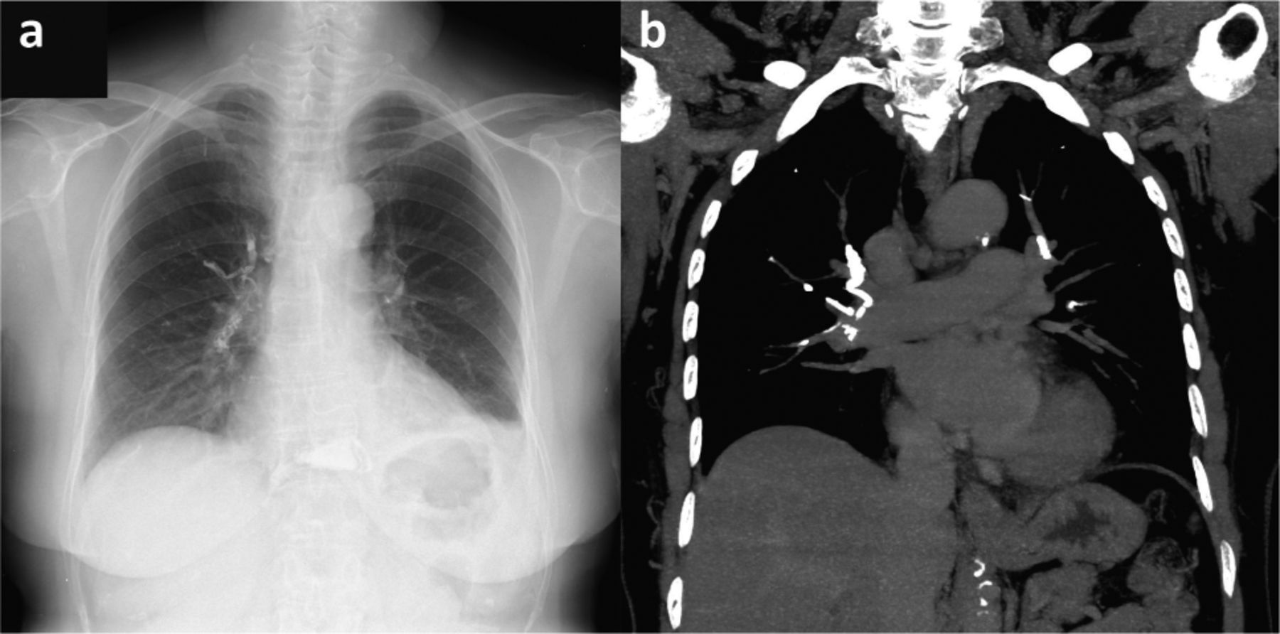
a) Plain chest X-ray showing high-density opacity in tubular branching pattern corresponding to pulmonary arterial distribution and left lower lobe consolidation. b) Coronal computed tomography of the thorax revealing segmental pulmonary cement embolism in both lungs.

Fig 2.
  • Download figure
  • Open in new tab
  • Download powerpoint
Fig 2.

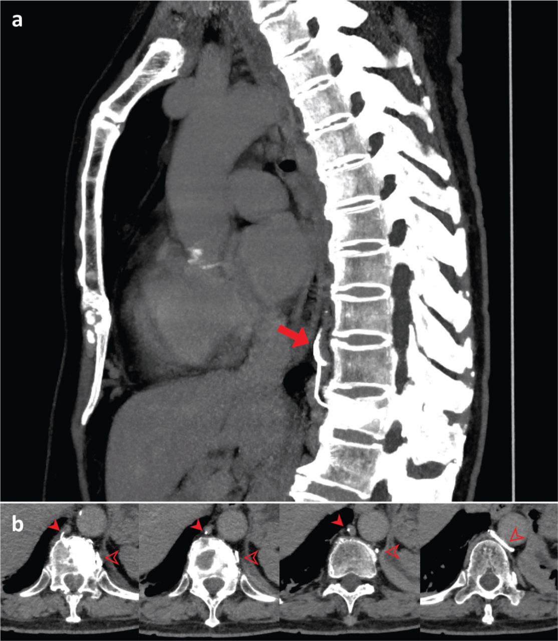
a) Sagittal computed tomography of the thorax demonstrates the cement leaking from T11 spine into the vertebral venous plexus (red arrow). b) Axial computed tomography showing evidence of cement leaking from PV site into the azygos (filled arrow) and hemiazygos (transparent arrow) venous system into pulmonary arterial circulation.

The patient subsequently underwent flexible bronchoscopy, which revealed purulent secretion only from the left lower lobe, with no foreign body, consistent with aspiration pneumonia. Her acute symptoms resolved after 2 weeks of antibiotics. She was managed conservatively for the central pulmonary cement embolism, as the event was years past, and she remained asymptomatic.

Discussion

PV involves injection of polymethylmethacrylate (PMMA) bone cement into the vertebral body for relief of pain and strengthening of bone in symptomatic vertebral compression fractures not feasible for definitive operation. Passage of bone cement into the vertebral venous plexus and then into the lungs is a rare but potentially serious complication of PV with the reported incidence up to 26%.1

Patients with cement embolism can present with a spectrum of symptoms – the diagnosis was picked up incidentally in our case when she presented with an unrelated aspiration pneumonia. No treatment is recommended for asymptomatic patients with peripheral cement embolisms. Surgical treatment with embolectomy is suggested for symptomatic central embolisms. Whereas, for symptomatic peripheral embolisms, or asymptomatic central embolisms as in our case, initial treatment should have been heparinisation followed by 3–6 months of warfarin therapy.2

This case highlights the need for a standard post-PV chest X-ray, as patients with cement embolisms can be completely asymptomatic. Timely recognition of her central event would have prompted initiation of anticoagulation. Measures to minimise the risk of pulmonary cement embolisms include avoidance of prone positioning, utilisation of fluoroscopy or computed tomography monitoring during PV procedure, and usage of bone cement in appropriate amount and viscosity.3

Acknowledgements

We thank our colleagues from Department of Diagnostic Imaging, Sarawak General Hospital, for a detailed analysis of the patient's medical imaging that greatly improved the manuscript.

  • © Royal College of Physicians 2021. All rights reserved.

References

  1. ↵
    1. Barbero S
    , Casorzo I, Durando M, et al. Percutaneous vertebroplasty: The follow-up. Radiol Med 2008;113:101–13.
    OpenUrl
  2. ↵
    1. Krueger A
    , Bliemel C, Zettl R, et al. Management of pulmonary cement embolism after percutaneous vertebroplasty and kyphoplasty: A systematic review of the literature. Eur Spine J 2009;18:1257–65.
    OpenUrlCrossRefPubMed
  3. ↵
    1. Baroud G
    , Crookshank M, Bohner M. High-viscosity cement significantly enhances uniformity of cement filling in vertebroplasty: an experimental model and study on cement leakage. Spine 2006;31:2562–8.
    OpenUrlCrossRefPubMed
Back to top
Previous articleNext article

Article Tools

Download PDF
Article Alerts
Sign In to Email Alerts with your Email Address
Citation Tools
Images of the month: Pulmonary cement embolism
Chan Sin Chai, Sze Shyang Kho, Swee Kim Chan, Teng Teng Tee, Siew Teck Tie
Clinical Medicine Jan 2021, 21 (1) e114-e115; DOI: 10.7861/clinmed.2020-0913

Citation Manager Formats

  • BibTeX
  • Bookends
  • EasyBib
  • EndNote (tagged)
  • EndNote 8 (xml)
  • Medlars
  • Mendeley
  • Papers
  • RefWorks Tagged
  • Ref Manager
  • RIS
  • Zotero
Share
Images of the month: Pulmonary cement embolism
Chan Sin Chai, Sze Shyang Kho, Swee Kim Chan, Teng Teng Tee, Siew Teck Tie
Clinical Medicine Jan 2021, 21 (1) e114-e115; DOI: 10.7861/clinmed.2020-0913
del.icio.us logo Digg logo Reddit logo Twitter logo Facebook logo Google logo Mendeley logo
  • Tweet Widget
  • Facebook Like
  • Google Plus One

Jump to section

  • Article
    • ABSTRACT
    • Case presentation
    • Discussion
    • Acknowledgements
    • References
  • Figures & Data
  • Info & Metrics

Related Articles

  • No related articles found.
  • PubMed
  • Google Scholar

Cited By...

  • No citing articles found.
  • Google Scholar

More in this TOC Section

  • Images of the month 1: Ischaemic stroke due to pulmonary arteriovenous fistula
  • Images of the month 1: Histoacryl glue embolisation to the right ventricle following treatment for gastric varices
  • Images of the month 2: Disseminated nocardiosis in a 60-year-old woman with sarcoidosis
Show more Image of the month

Similar Articles

FAQs

  • Difficulty logging in.

There is currently no login required to access the journals. Please go to the home page and simply click on the edition that you wish to read. If you are still unable to access the content you require, please let us know through the 'Contact us' page.

  • Can't find the CME questionnaire.

The read-only self-assessment questionnaire (SAQ) can be found after the CME section in each edition of Clinical Medicine. RCP members and fellows (using their login details for the main RCP website) are able to access the full SAQ with answers and are awarded 2 CPD points upon successful (8/10) completion from:  https://cme.rcplondon.ac.uk

Navigate this Journal

  • Journal Home
  • Current Issue
  • Ahead of Print
  • Archive

Related Links

  • ClinMed - Home
  • FHJ - Home
clinmedicine Footer Logo
  • Home
  • Journals
  • Contact us
  • Advertise
HighWire Press, Inc.

Follow Us:

  • Follow HighWire Origins on Twitter
  • Visit HighWire Origins on Facebook

Copyright © 2021 by the Royal College of Physicians