
ABSTRACT
Atopic dermatitis (AD) is a common inflammatory skin disease characterised by itch and is responsible for significant reduction in quality of life. While AD primarily arises in those under the age of 2 years, it is frequently persistent into adulthood. Recognition of AD is important for the general physician, especially to distinguish causes of acute flares that may present in any medical setting, such as eczema herpeticum and associated allergic reactions. While, to date, treatments have largely focused on broad spectrum immunomodulation with corticosteroids or systemic therapies (such as ciclosporin and methotrexate), increased knowledge in the pathophysiology of the disease has recently led to the expansion of treatment options available for those suffering with AD, and the new drugs on the horizon promise a previously unimagined potential for effective and safe treatment.
Key points
Atopic dermatitis (AD) is a common itchy skin condition occurring in those with a genetic tendency and has the potential to be significantly debilitating to both children and adults.
Regular application of topical emollients alongside treatment with topical corticosteroids or calcineurin inhibitors forms the foundation of treatment of all AD.
Flares of AD may be driven by secondary infection with bacteria (eg Staphylococcus aureus) or virus (eg herpes simplex virus (eczema herpeticum)) and require same-day dermatological advice.
Patients may present to the general medic with co-existing AD, and prescribing of other therapies such as oral corticosteroids may need to be adjusted for the presence of eczema (eg treatment tapered slowly).
New treatments for AD are dramatically changing the treatment expectations for patients and doctors because of superior efficacy and safety. These new therapies target the consequences of T cell activation including Th2 cytokines (eg dupilumab (blocks IL-4/IL-13 receptor)) and multiple cytokine pathways (eg Janus kinase (JAK) inhibitors (baracitinib, abrocitinib and upadacitinib)).
Introduction
Atopic dermatitis (AD) is one of the most common chronic inflammatory skin diseases in the UK, responsible for a large proportion of consultations in both primary and secondary care in the acute and outpatient setting. Dermatitis (inflammation of the skin) is a term used synonymously with eczema and atopic eczema is synonymous with AD. Here, we discuss AD, for which there are clear diagnostic criteria (Box 1).1 Individuals with AD show a genetic predisposition to allergic conditions (such as asthma, allergic rhinitis, allergic conjunctivitis and food allergy), but demonstration of allergic responses (eg measurement of specific immunoglobulin (Ig) E) are not required to make the diagnosis. Importantly, in the dermatological lexicon, the term ‘eczema’ is overarching and, as well as AD, also encompasses other forms (such as contact irritant, contact allergic, asteototic and venous).2,3
Diagnostic criteria of atopic dermatitis (eczema)1
AD can affect any age but is predominantly a disease of childhood and is estimated to affect up to 30% of UK school children. Although epidemiological studies vary, approximately 10% of adults are affected. Adult onset AD (late onset) is also recognised.2,4 While many cases of AD are mild, and can be managed with simple topical treatment, up to 50% of AD cases are moderate–severe and these individuals suffer considerable loss of quality of life caused by the impact of chronic itching, loss of sleep, painful skin and social isolation due to low self-esteem.
How to recognise eczema
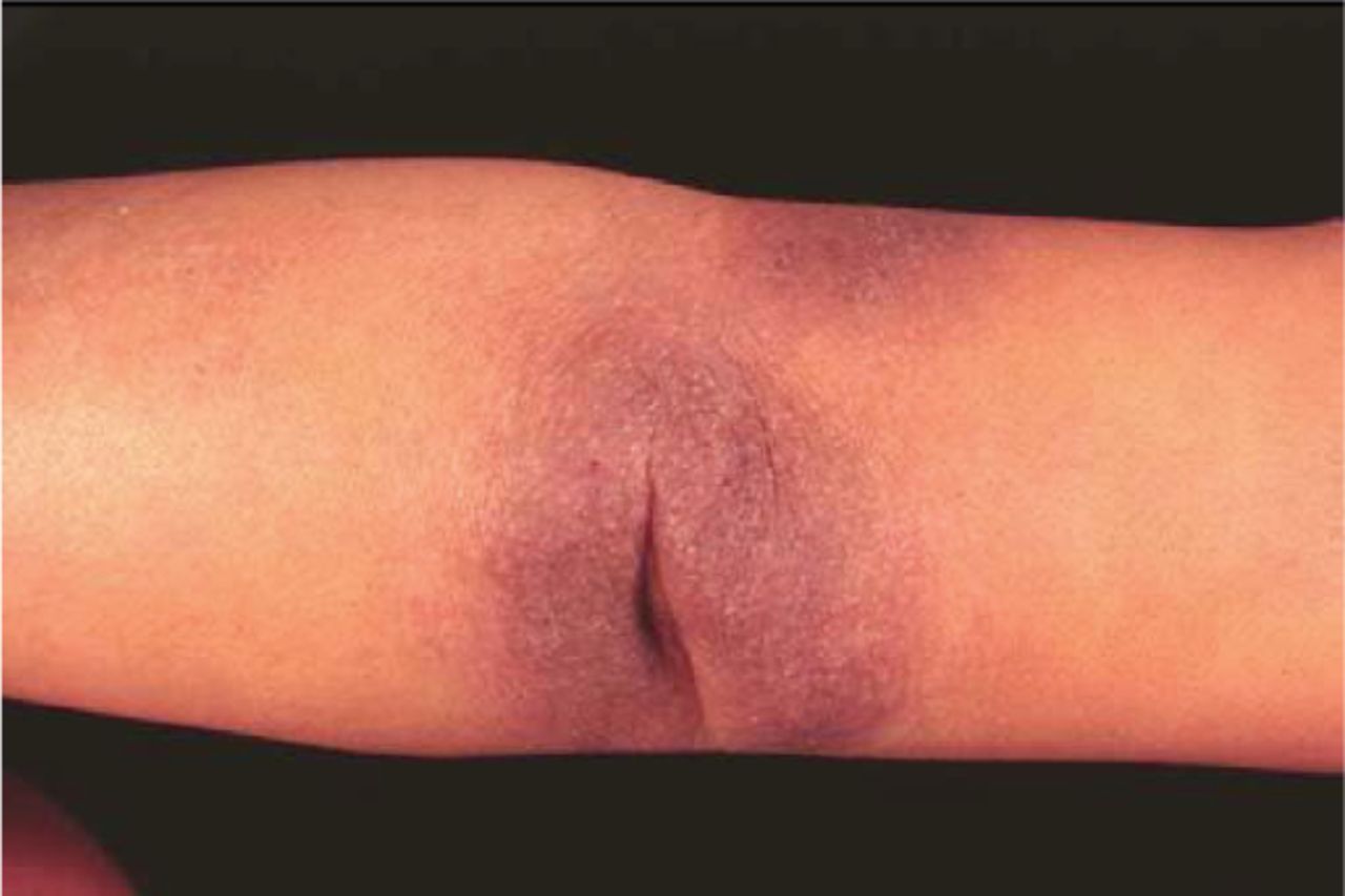
Diagnostic criteria were developed for clinical trials and provide a simple approach to diagnosis (Box 1). The critical symptom of AD is itch, which can be debilitating. Clinical findings are characterised by ill-defined, papular erythema and fine scale, typically affecting the flexures (antecubital and popliteal fossae, neck, wrists, ankles and eyelids). Chronic eczema results in thickening and increased scaling of the skin (lichenification; Fig 1). Importantly, in dark skin, the erythema is often not visible and instead the scaling or thickening from lichenification may be more prominent with hyper- or hypopigmentation (Fig 2). Generalised dry skin (xerosis and/or ichthyosis vulgaris) is typical in more extensive cases. Depending on the severity and chronicity of disease, skin findings may also include exudate, vesicles, fissures, lichenification and excoriations.1–3,5 In dark skin, temporary post-inflammatory changes to skin pigmentation may arise after treatment and can cause either darkening or lightening. When skin disease is severe, erythroderma can occur, where more than 90% of the body surface area is affected.
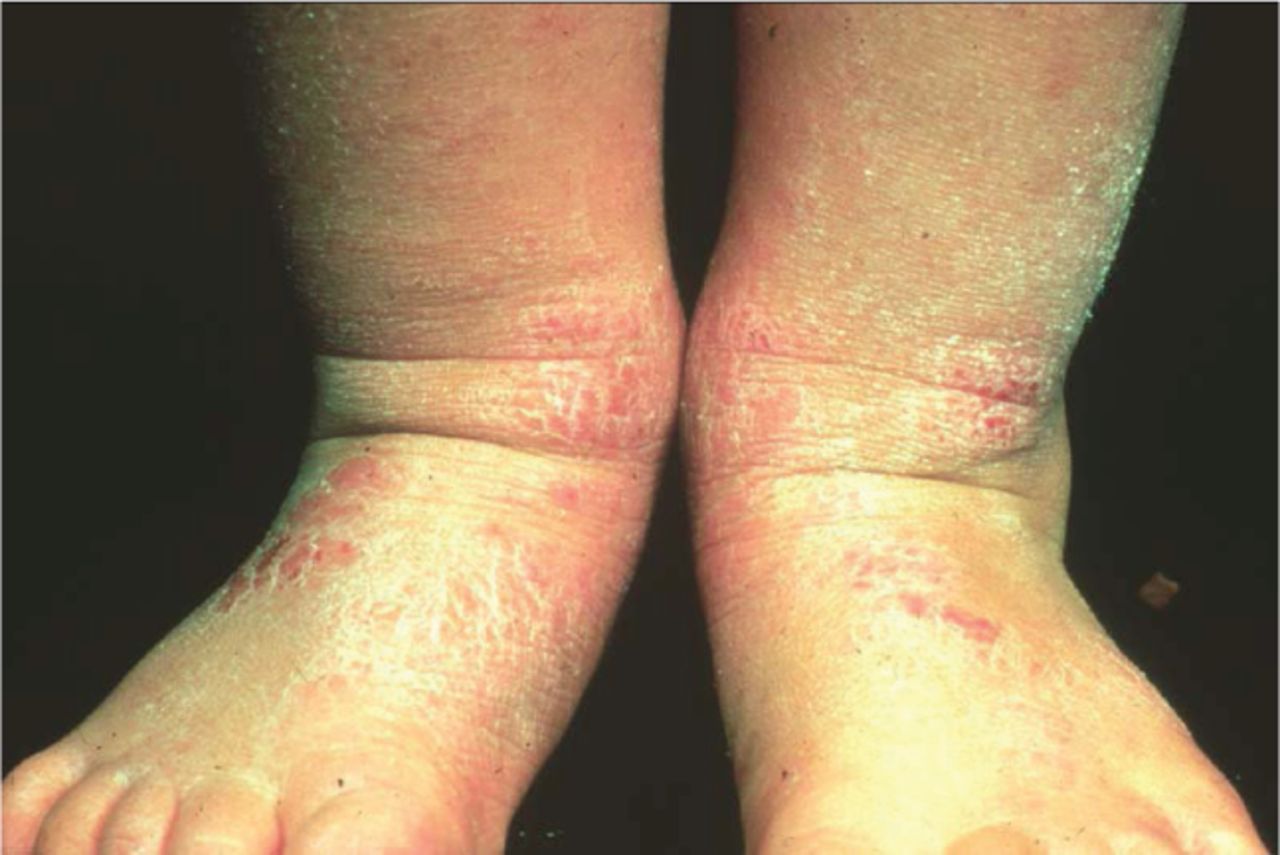
Lichenified (thickened), scaly skin over flexural sites of chronic eczema. Inflammation is evident.

Lichenified skin at a site of chronic eczema in dark skin, showing hyperpigmentation. Inflammation is not evident.
Complications of AD include skin infection with bacteria (commonly Staphylococcus aureus), viruses (especially disseminated herpes simplex/eczema herpeticum) and yeasts. Individuals with AD have an increased risk of various atopic conditions including asthma, allergic rhinitis, allergic keratoconjunctivitis and food allergy. Other autoimmune conditions such as alopecia areata are also more prevalent.
Pathogenesis
Patients with AD have a genetic predisposition to a disrupted skin barrier and a propensity to make aberrant cutaneous immune responses. The former is demonstrated by increased permeability of the epidermis to water (resulting in dry skin) and the latter by a specific predisposition to development of type 2 immunity as a consequence of exposure to multiple antigens in the skin. The precise link between the two remains unclear, but the concept of the disrupted barrier providing the increased allergen exposure and subsequent Th2 primed immune responses accounting for the associated allergic sensitivity is widely accepted.2,3,6,7 Importantly, there is no evidence for an altered systemic immune response in AD, although rarely specific primary immunodeficiencies (such as hyper-IgE syndrome) can present with atopic dermatitis-like features. Genome-wide association studies led to the discovery of multiple genetic loci for AD susceptibility, but the most replicated and strongest of all associations is with the gene encoding filaggrin (FLG). Subsequently, loss-of-function FLG mutations have been identified in populations across the globe and are associated with increased risk of AD. FLG mutations, and are carried in approximately 10% of all Europeans, and individuals carrying these show an increased risk of development of AD (odds ratio (OR) 3.39–4.78). Filaggrin is a skin barrier protein expressed in the outer layers of the epidermis and all mutations reduce expression, and are associated with a more severe phenotype and early onset disease that persists into adulthood.
Regarding allergy and AD, there are commonly misunderstood, important concepts in AD pathogenesis. AD is not an allergy per se, instead, AD inflammation is mediated by T cell activation in the skin where the antigenic targets for activation are likely to be numerous in any one individual. IgE is considered to play little in the role of mediating the skin inflammation, yet, in individuals with AD, IgE allergies are more common and account for the association with allergic rhinitis, food allergy and asthma. Induction of IgE responses in AD is thought to be a consequence of increased allergen exposure through broken skin in the setting of a cutaneous drive towards the initiation of Th2 responses (which subsequently induce B cell class switching to IgE production). However, while the primary skin inflammation of AD is not IgE driven, recurrent allergic reactions causing mast cell degranulation with wheal-and-flare reactions (urticaria) in the skin will exacerbate AD through increased inflammation and scratching. As a consequence of the increased propensity to induction of specific IgE in AD, allergic reactions to foods are identified in approximately 30% of children with moderate–severe AD. However, food allergic reactions in adults with AD are much less prevalent. Most children with AD and food allergy have a history of immediate reactions following ingestion of the food in question (within 1 hour) and, therefore, these foods should be strictly avoided until further specialist paediatric allergy advice (for fear of risking subsequent anaphylaxis). For children without immediate reactions, food is unlikely to be a cause of AD exacerbation but this should be considered if response to treatment is poor; specialist advice is recommended. Aeroallergens are not a common cause for flares of AD.
Therefore, investigation of all cases for allergies with a ‘shotgun’ approach is not indicated. Instead, for all patients with AD, exploration of allergy should be undertaken by a focused history and allergic triggers should be followed up by specialist advice. Importantly, while most IgE allergies can be confirmed by skin prick testing or specific IgE measurement, these are an unreliable means to screen for an allergy relevant to non-immediate reactions and specialist dermatological advice is recommended.
Why is it important?
The burden of AD is far reaching, with significant impact on quality of life of affected patients and their household. Severe itching causes chronic sleep disturbance, impacting on child growth and development and neurocognitive ability for both children and adults alike. This sleep disturbance, coupled with intense pruritus and laborious topical treatment regimens, results in days off work and impacts relationships. This is reflected in the Global Burden of Disease study, showing that dermatitis was the leading skin disease, measured by disability-adjusted life-years.4,8,9
Patients with AD are more likely to require treatment for associated atopic comorbidities (such as asthma, allergic rhinitis and type-1 hypersensitivity to food), therefore, suffering an even greater disease burden.9 Increased prevalence of depression and anxiety is also seen.9,10 Furthermore, uncontrolled moderate–severe disease is associated with increased cardiovascular risk.9
Managing AD
For current principles of atopic dermatitis management, see Fig 4.
AD for the general medic
Due to the prevalence of AD it is highly likely the general medic will encounter affected patients. Patients may present acutely with flares of their AD resulting in systemic upset and/or erythroderma. Reasons for an acute exacerbation may be unclear but it is important to consider infection, commonly as a result of increases in density of S aureus. Treatment of any suspected bacterial infection should be given in tandem with topical AD treatment (usually topical corticosteroids). Eczema herpeticum (EH; disseminated cutaneous herpes simplex virus) is a severe complication and must be dealt with urgently with referral to dermatology. EH is typically painful and characterised clinically by clusters of monomorphic vesicles (equal sized small blisters) which can become crusted leaving a ‘punched out appearance’ (Fig 3). EH is more common in children or those on immunosuppressants and necessitates prompt treatment with systemic antiviral treatment (aciclovir or valaciclovir). Due to the risk of corneal ulceration, periorbital involvement requires urgent ophthalmological review.2,3
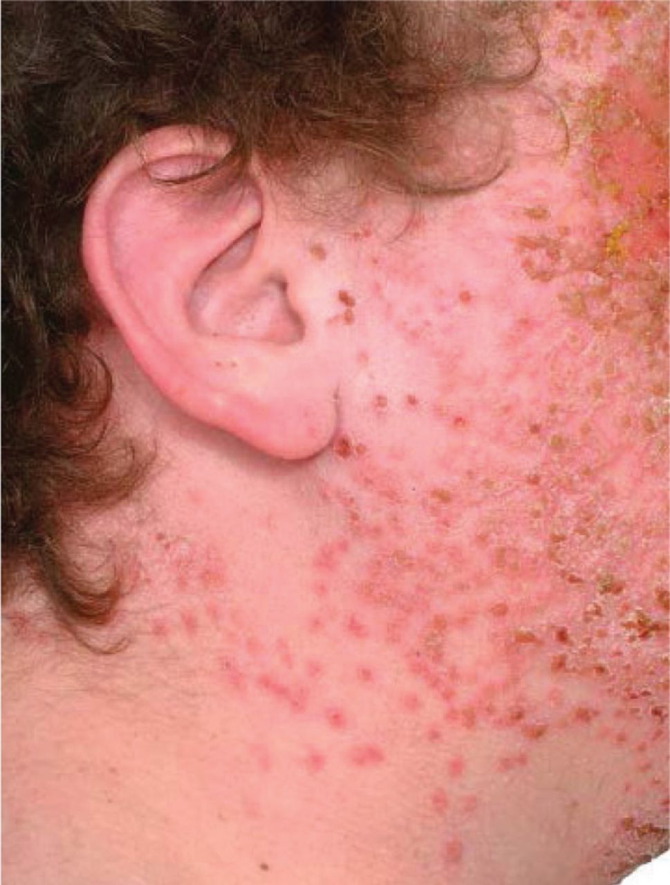
Diffuse spread of small vesicles across the face with crusting and scaling in a patient with atopic dermatitis.
Patients with AD may also present due to their atopic comorbidities or due to unrelated disease. As inpatients, concurrent illness, addition of new medications and irritancy from detergent-containing washes can flare AD. It is frequently observed that AD treatments may be missed from inpatient prescribing, leading to skin deterioration. Interestingly, concomitant benefit to AD from oral prednisolone may be seen during treatment for other conditions (eg flares of asthma), but abrupt withdrawal usually induces a flare in the skin disease worse than the original eczema severity, necessitating gentle tapering of oral corticosteroids over 4–6 weeks in those with moderate–severe AD. For AD alone, almost any severity can be overcome during inpatient stay with intensive topical treatment alone. Therefore, oral corticosteroids are generally not recommended outside of dermatological advice.
Furthermore, it is important for the general medic to recognise the potential complications of immunosuppression in those individuals on systemic medications and recognise the specific risks of the targeted agents increasingly available (detailed later).
Measuring disease severity
Various objective measures of severity (Eczema Area and Severity Index (EASI)) and measures of subjective severity by questionnaire (Dermatology Life Quality Index (DLQI) or Patient Orientated Eczema Management (POEM)) are validated for use in adults and children and are increasingly part of dermatological practice. It is likely these read-outs will become widespread in primary and secondary care for guidance on effective management and the options available for treatment.
Advances in treatment
As shown in Fig 4, topical emollients and anti-inflammatories form the foundation of treatment for AD.2,3,5 A significant proportion of patients with moderate–severe disease are refractory to topical therapy. In current practice, these patients are escalated to phototherapy or systemic immunosuppression such as ciclosporin, methotrexate, azathioprine and mycophenolate mofetil. Since 2018, dupilumab, the first biologic agent available for AD, has transformed current practice.11,12
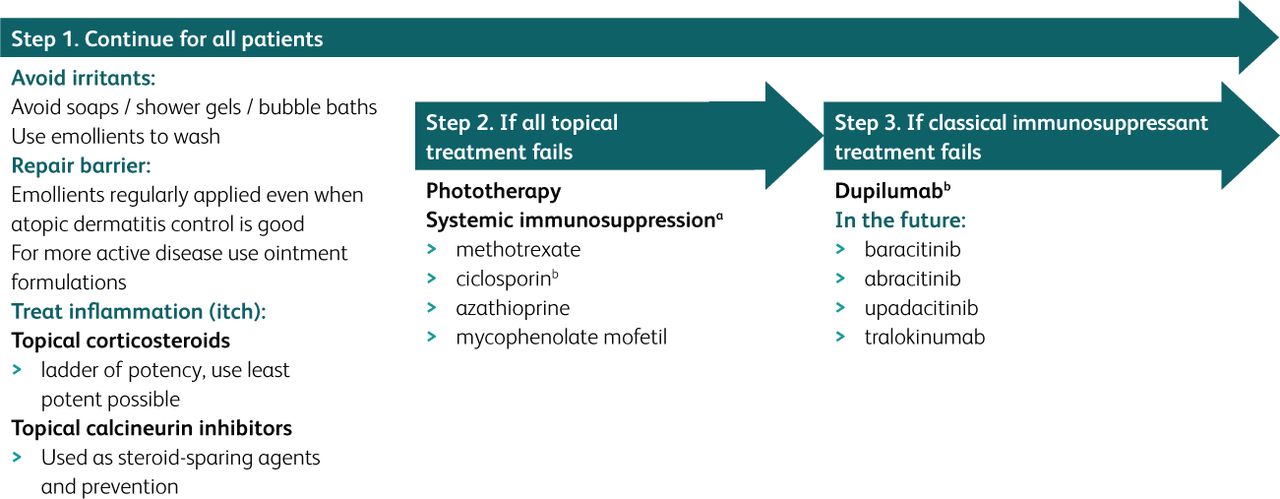
Current principles of atopic dermatitis management. aAccording to National Institute for Health and Care Excellence guidance, failure of one systemic immunosuppressant is required before proceeding to step 3; bciclosporin and dupilumab are the only licensed systemic therapies for atopic dermatitis.
Interleukin-4 and/or interleukin-13 inhibition (dupilumab and tralokinumab)
Advancements in our understanding of the pathophysiology of AD and chronic itch have led to the development of new therapeutic targets and, excitingly, novel therapies are slowly revolutionising its treatment. In 2016, the publication of two randomised, placebo-controlled trials (SOLO1 and 2) comparing dupilumab (a monoclonal antibody targeting the interleukin (IL)-4/IL-13 receptor) with placebo showed promising results at 16 weeks’ treatment with reduction in global assessment scores, AD severity scores, itch scores as well as improvement in anxiety, depression and quality of life. In 2018, the National Institute for Health and Care Excellence (NICE) approved the use of dupilumab in patients with moderate–severe AD who had not responded to at least one systemic therapy and it was later approved for the use in children over the age of 12 years.11–14
The most frequent adverse effects of dupilumab are injection site reactions and non-infectious conjunctivitis, which rarely necessitates treatment cessation. Risk of serious infection with dupilumab appears low and biologics targeting the Th2 pathway do not significantly increase bacterial or opportunistic infections. Viral infections (mucosal herpes virus and nasopharyngitis) appear to be increased but conversely dupilumab may have a protective effect against cutaneous viral infections. Dupilumab should be avoided in patients at risk of helminth infections.14,15 Tralokinumab (anti-IL-13) has nearly completed phase III clinical trials and early reports have shown that, as well as dupilumab, alternative strategies for Th2 inhibition by direct anti-cytokine monoclonal antibody therapy with anti-IL-13 are likely to be effective.
Janus kinase inhibitors (including baracitinib, abrocitinib and upadacitinib)
Following the demonstration by dupilumab that blockade of the IL-4 and IL-13 pathway is effective in the management of AD, and the knowledge that other inflammatory pathways are also active in the skin, blockade of the intracellular pathway from multiple cytokine receptors by the shared Janus kinase (JAK) – signal transducer and activator of transcription proteins (STAT) pathway has been seen as an attractive target in AD.16,17 JAKs are an important signalling pathway for multiple cellular processes including haematopoiesis. Hence, earlier drug designs with broad activity were limited by their adverse events profiles. Recent work to identify selectivity for specific molecules to certain JAK isoforms (JAK inhibitors (JAKis)) has shown that specific cytokine receptors can be targeted, and also shown that the JAKi adverse event profile can be improved. This has facilitated design of JAKis for a variety of inflammatory diseases (including psoriasis, inflammatory bowel disease and rheumatoid arthritis) with therapies bespoke to the key pathways at work. In addition, tissue specific effects of JAKs may also be targeted. For example, JAK1 is expressed in itch sensory neurons and higher selectivity against JAK1 may be associated with a favourable benefit–risk profile.16–19 JAKis are oral medications, and therefore offer alternative treatment options for patients unable to receive injections.
Baracitinib, an oral JAK inhibitor (JAK1 and JAK2) was shown in BREEZE-AD7, a randomised placebo-controlled phase III trial, to improve disease severity and itch scores and is now licensed for use in atopic dermatitis but is yet to be approved by NICE.20 It is already indicated in patients with rheumatoid arthritis who have failed disease-modifying antirheumatic drugs (DMARD) therapy.
Data from two phase III trials (JADE MONO-1 and -2) comparing abrocitinib (an oral selective JAK1 inhibitor) to placebo included adults and adolescents over the age of 12. Sixty-three per cent and 61% of patients receiving 200 mg abrocitinib monotherapy vs 12% and 10% of those in the placebo groups (p<0.01) achieved a 75% reduction in their AD severity score at 12 weeks.18 A comparison study of abrocitinib, dupilumab and placebo, with all subjects applying a standardised topical therapy regimen throughout, suggests abrocitinib may provide a more rapid reduction in itch.17 In February 2021, the Medicines and Healthcare products Regulatory Agency (MHRA) issued early access to abrocitinib in the early access to medicines scheme.
Upadacitinib, another JAK1 inhibitor approved for use in rheumatoid arthritis, also shows promising results from preliminary phase III trial data in AD. At 16 weeks, a 75% improvement in AD severity score was seen in 80% of patients receiving upadacitinib 30 mg once daily monotherapy compared with 16% in the placebo group (p<0.001). Adolescents were included in the trials and showed a similar response to treatment.19,20
Adverse effects with JAK inhibition appears to include a slightly increased frequency of infections, especially upper respiratory tract infections and reactivation of herpes zoster and herpes simplex. Thromboembolic events are rare and have not shown a strong signal in preclinical testing, but real-world data will define whether these are a significant caution. All JAKi treatments should be discontinued if clinical features of deep vein thrombosis/pulmonary embolism occur. Most JAKis require monitoring for haematological changes (thrombocytopenia), more so if JAK2 is inhibited, and blood lipid monitoring is recommended. The dose of JAKis should be reduced in patients receiving strong inhibitors of cytochrome P450.17–20
Other
Other areas of research include targeted therapies to additional inflammatory cytokines or their receptors, such as IL-31, IL-33 and thymic stromal lymphopoietin (TSLP), with several phase II and III trials underway.16 Increasingly, with progression in gut microbiome research, attention is drawn to the skin microbiome and its implication in skin disease. Overgrowth of S aureus in lesional skin of AD patients has long been recognised. This is a complex interaction with, as yet, only small single studies suggesting distinct microbial communities present in the nose and skin of affected individuals.21
Conclusion
AD, a common dermatological diagnosis with significant disease burden and impact on patient quality of life, has long been limited with its treatment armamentarium. Management of moderate–severe disease has resulted in the use of immunosuppressive and potentially toxic drugs, often in young populations. New developments in the understanding of its pathophysiology and, hence, targeted biologic therapy show a promising stride towards addressing an unmet need in AD treatment.
Conflicts of interest
Michael Ardern-Jones has been as a consultant, advisor, speaker, grant holder or collaborator (academic) for Pfizer, Sanofi-Genzyme, Leo Pharma, UCB, Hosei-Sptares, Ducentis, AbbVie, Amgen and Unilever.
- © Royal College of Physicians 2021. All rights reserved.


