
Main menu

  • Home
  • Our journals
    • Clinical Medicine
    • Future Healthcare Journal
  • Subject collections
  • About the RCP
  • Contact us

Clinical Medicine Journal

  • ClinMed Home
  • Content
    • Current
    • Ahead of print
    • Archive
  • Author guidance
    • Instructions for authors
    • Submit online
  • About ClinMed
    • Scope
    • Editorial board
    • Policies
    • Information for reviewers
    • Advertising

User menu

  • Log in

Search

  • Advanced search
RCP Journals
Home
  • Log in
  • Home
  • Our journals
    • Clinical Medicine
    • Future Healthcare Journal
  • Subject collections
  • About the RCP
  • Contact us
Advanced

Clinical Medicine Journal

clinmedicine Logo
  • ClinMed Home
  • Content
    • Current
    • Ahead of print
    • Archive
  • Author guidance
    • Instructions for authors
    • Submit online
  • About ClinMed
    • Scope
    • Editorial board
    • Policies
    • Information for reviewers
    • Advertising

Images of the month 2: A target sign in COVID-19 pneumonia

Jitender Singh and Tarika Sharma
Download PDF
DOI: https://doi.org/10.7861/clinmed.2021-0377
Clin Med July 2021
Jitender Singh
AShanti Mukund Hospital, New Delhi, India
Roles: consultant
  • Find this author on Google Scholar
  • Find this author on PubMed
  • Search for this author on this site
  • For correspondence: introductory2008@gmail.com
Tarika Sharma
BCollege of Nursing at the Institute of Liver and Biliary Sciences, New Delhi, India
Roles: assistant professor
  • Find this author on Google Scholar
  • Find this author on PubMed
  • Search for this author on this site
  • Article
  • Figures & Data
  • Info & Metrics
Loading

ABSTRACT

A target sign has not been described in other viral or bacterial pneumonia on computed tomography of the chest in literature. It could represent a hallmark of COVID-19 pneumonia, given the good correlating clinical context and prevalence of COVID-19.

KEYWORDS:
  • computed tomography
  • reverse transcription polymerase chain reaction
  • COVID-19, ground-glass opacifications
  • target sign

Case presentation

A 37-year-old man presented with fever of 5 days duration. At the time of admission, the patient had breathlessness along with hypoxia, reverse transcription polymerase chain reaction was positive. X-ray of the chest revealed small patchy ground-glass haziness in bilateral lower lung fields (right>left). The patient was shifted to the high dependency unit due to worsening of respiratory symptoms on day 9 and a repeat computed tomography (CT) was performed which showed consolidation and ground-glass opacification (GGO) along with a target sign in subpleural location of right lower lung lobe (Fig 1a). He was managed with oxygen therapy and other medications. The patient improved and was discharged in a stable condition on day 17.

Fig 1.
  • Download figure
  • Open in new tab
  • Download powerpoint
Fig 1.

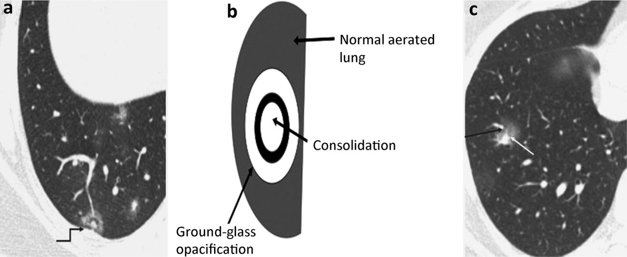
Target sign and halo sign. a) Computed tomography (coned down axial section) of the right lung showing an irregular ring-like opacity containing in its centre, a small nodular ground-glass opacification surrounding a very small vessel (arrow); also of note are small patchy ground-glass opacities. b) Graphic showing how to identify a target sign. c) Computed tomography (coned down axial section at cardiac level) showing combination of central nodular opacity and the peripheral irregular rim of ground-glass opacification; also of note are small patchy ground-glass opacities.

Discussion

The sign was first described in June 2020 in COVID-19 pneumonia on CT of the chest.1

The sign has been depicted as a central high attenuation focus surrounded by one or more dense complete or incomplete ring-like consolidations, forming one or more circles on CT of the chest in patients with COVID-19 pneumonia (Fig 1b).1 The pathophysiological sign is a manifestation of organising pneumonia (OP) which represents peripheral opacities whereas the central nodular opacity represents the perivascular inflammation or focal enlargement of the pulmonary artery (vessel sign).2,3 The pulmonary target sign usually coexists with other typical OP features including the reverse halo or halo sign which are characterised by peripheral consolidation with central GGO and central consolidation with peripheral GGO, respectively (Fig 1c).4 Further patho-radiological studies are required to investigate the value of this sign in the specificity with COVID-19 pneumonia.

  • © Royal College of Physicians 2021. All rights reserved.

References

  1. ↵
    1. Müller CIS
    , Müller NL. Chest CT target sign in a couple with COVID-19 pneumonia. Radiol Bras 2020;53:252–4.
    OpenUrl
  2. ↵
    1. Pogatchnik BP
    , Swenson KE, Sharifi H, et al. Radiology-pathology correlation in recovered COVID-19, demonstrating organizing pneumonia. Am J Respir Crit Care Med 2020;202:598–9.
    OpenUrl
  3. ↵
    1. Saburi A
    , Schoepf UJ, Ulversoy KA, et al. From radiological manifestations to pulmonary pathogenesis of COVID-19: a bench to bedside review. Radiol Res Pract 2020;2020:8825761.
    OpenUrl
  4. ↵
    1. Lang M
    , Som A, Carey D, et al. Pulmonary vascular manifestations of COVID-19 pneumonia. Radiol Cardiothorac Imaging 2020;2:e200277.
    OpenUrl
Back to top
Previous articleNext article

Article Tools

Download PDF
Article Alerts
Sign In to Email Alerts with your Email Address
Citation Tools
Images of the month 2: A target sign in COVID-19 pneumonia
Jitender Singh, Tarika Sharma
Clinical Medicine Jul 2021, 21 (4) e410-e411; DOI: 10.7861/clinmed.2021-0377

Citation Manager Formats

  • BibTeX
  • Bookends
  • EasyBib
  • EndNote (tagged)
  • EndNote 8 (xml)
  • Medlars
  • Mendeley
  • Papers
  • RefWorks Tagged
  • Ref Manager
  • RIS
  • Zotero
Share
Images of the month 2: A target sign in COVID-19 pneumonia
Jitender Singh, Tarika Sharma
Clinical Medicine Jul 2021, 21 (4) e410-e411; DOI: 10.7861/clinmed.2021-0377
del.icio.us logo Digg logo Reddit logo Twitter logo Facebook logo Google logo Mendeley logo
  • Tweet Widget
  • Facebook Like
  • Google Plus One

Jump to section

  • Article
    • ABSTRACT
    • Case presentation
    • Discussion
    • References
  • Figures & Data
  • Info & Metrics

Related Articles

  • No related articles found.
  • PubMed
  • Google Scholar

Cited By...

  • No citing articles found.
  • Google Scholar

More in this TOC Section

  • Images of the month 2: Extensive pneumocephalus after subdural haematoma
  • Images of the month 1: Trident sign and neurosarcoidosis
  • Images of the month 1: ‘Soy sauce’ pleural effusion: what causes black pleural fluid?
Show more Images of the month

Similar Articles

FAQs

  • Difficulty logging in.

There is currently no login required to access the journals. Please go to the home page and simply click on the edition that you wish to read. If you are still unable to access the content you require, please let us know through the 'Contact us' page.

  • Can't find the CME questionnaire.

The read-only self-assessment questionnaire (SAQ) can be found after the CME section in each edition of Clinical Medicine. RCP members and fellows (using their login details for the main RCP website) are able to access the full SAQ with answers and are awarded 2 CPD points upon successful (8/10) completion from:  https://cme.rcplondon.ac.uk

Navigate this Journal

  • Journal Home
  • Current Issue
  • Ahead of Print
  • Archive

Related Links

  • ClinMed - Home
  • FHJ - Home
clinmedicine Footer Logo
  • Home
  • Journals
  • Contact us
  • Advertise
HighWire Press, Inc.

Follow Us:

  • Follow HighWire Origins on Twitter
  • Visit HighWire Origins on Facebook

Copyright © 2021 by the Royal College of Physicians