
Main menu

  • Home
  • Our journals
    • Clinical Medicine
    • Future Healthcare Journal
  • Subject collections
  • About the RCP
  • Contact us

Clinical Medicine Journal

  • ClinMed Home
  • Content
    • Current
    • Ahead of print
    • Archive
  • Author guidance
    • Instructions for authors
    • Submit online
  • About ClinMed
    • Scope
    • Editorial board
    • Policies
    • Information for reviewers
    • Advertising

User menu

  • Log in

Search

  • Advanced search
RCP Journals
Home
  • Log in
  • Home
  • Our journals
    • Clinical Medicine
    • Future Healthcare Journal
  • Subject collections
  • About the RCP
  • Contact us
Advanced

Clinical Medicine Journal

clinmedicine Logo
  • ClinMed Home
  • Content
    • Current
    • Ahead of print
    • Archive
  • Author guidance
    • Instructions for authors
    • Submit online
  • About ClinMed
    • Scope
    • Editorial board
    • Policies
    • Information for reviewers
    • Advertising

Images of the month 3: Transient perivascular inflammation of the carotid artery syndrome

Philipp Jud, Georg Kangler, Paul Gressenberger, Rupert H Portugaller and Marianne Brodmann
Download PDF
DOI: https://doi.org/10.7861/clinmed.2021-0349
Clin Med July 2021
Philipp Jud
AMedical University of Graz, Graz, Austria
Roles: registrar angiologist
  • Find this author on Google Scholar
  • Find this author on PubMed
  • Search for this author on this site
  • For correspondence: philipp.jud@medunigraz.at
Georg Kangler
BMedical University of Graz, Graz, Austria
Roles: registrar otorhinolaryngologist
  • Find this author on Google Scholar
  • Find this author on PubMed
  • Search for this author on this site
Paul Gressenberger
CMedical University of Graz, Graz, Austria
Roles: registrar angiologist
  • Find this author on Google Scholar
  • Find this author on PubMed
  • Search for this author on this site
Rupert H Portugaller
DMedical University of Graz, Graz, Austria
Roles: consultant radiologist
  • Find this author on Google Scholar
  • Find this author on PubMed
  • Search for this author on this site
Marianne Brodmann
EMedical University of Graz, Graz, Austria
Roles: consultant angiologist
  • Find this author on Google Scholar
  • Find this author on PubMed
  • Search for this author on this site
  • Article
  • Figures & Data
  • Info & Metrics
Loading
KEYWORDS:
  • carotid artery
  • vascular disease

Case presentation

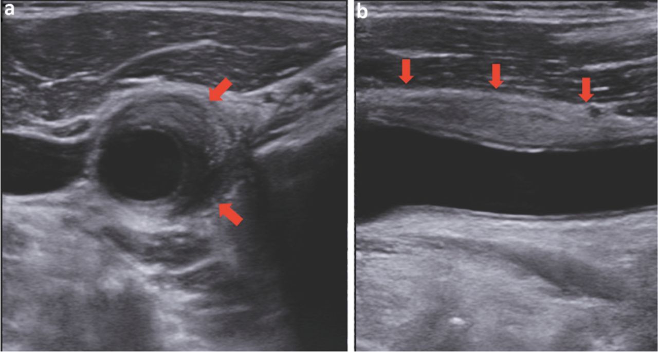
A 44-year-old man presented to the emergency department with a painful, palpable mass in the area of the right carotid triangle lasting for 1 week. He was otherwise healthy without any trauma or further otorhinolaryngological symptoms. Other than a pain at the right carotid triangle on pressure, physical and otorhinolaryngological examinations were unremarkable. Ultrasonography of the neck revealed a hypoechogenic mass in the adventitial layer of the right medial common carotid artery with a crescent shape in transverse view (Fig 1a) reaching from the distal common carotid artery to the carotid bulb (35 × 12 × 6 mm; Fig 1b). Carotid intima-media thickness was normal and no dissection membrane could be found. Subsequent ultrasonography of the aorta, its major branches and temporal arteries was also unremarkable, while magnetic resonance imaging of the carotid arteries revealed local contrast enhancement of the right common carotid artery (Fig 2). No potential signs or symptoms for inflammation, especially vasculitis, were found and comprehensive laboratory testing including inflammatory parameters, thyroid hormones, autoantibodies and immunoglobulin G4 was unremarkable. 18-fluorodeoxyglucose positron emission tomography – computed tomography showed only an increased tracer uptake of the right common carotid artery. Diagnosis of transient perivascular inflammation of the carotid artery (TIPIC) syndrome was made and dexibuprofen 400 mg twice daily together with pantoprazole 40 mg once daily peroral were prescribed for 2 weeks. The pain resolved within a few days and the hypoechogenic mass was undetectable in a follow-up visit 2 weeks later. Since the patient's initial presentation, he was still asymptomatic 1 year later.

Fig 1.
  • Download figure
  • Open in new tab
  • Download powerpoint
Fig 1.

Ultrasonography of the neck showing a hypoechogenic mass in the adventitial layer of the right medial common carotid artery with a crescent shape in transverse view reaching from the distal common carotid artery to the carotid bulb.

Fig 2.
  • Download figure
  • Open in new tab
  • Download powerpoint
Fig 2.

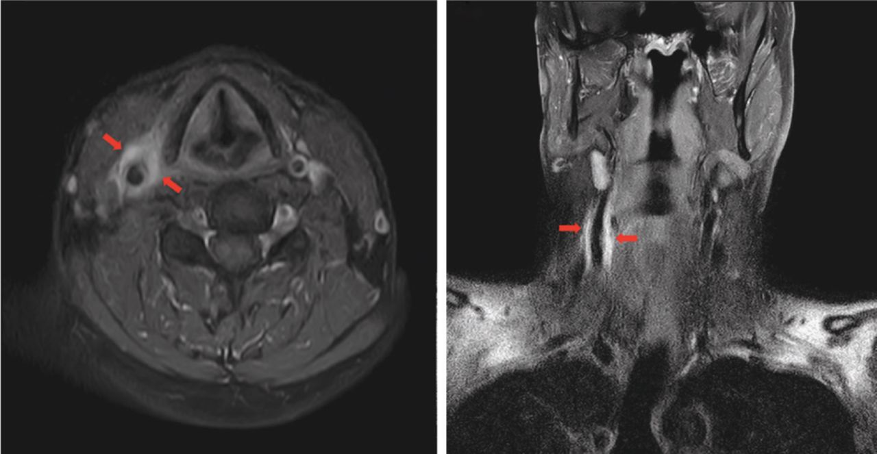
Magnetic resonance imaging of the carotid arteries showing local contrast enhancement of the right common carotid artery.

Discussion

TIPIC syndrome, formerly known as idiopathic carotidynia, was firstly described by Fay in 1927 and recent data suggested that idiopathic carotidynia is a distinct disease due to imaging abnormalities of the carotid artery and introduced the term ‘transient perivascular inflammation of the carotid artery syndrome’.1,2 TIPIC syndrome is a rare, clinicoradiologic entity with crescent thickening of the carotid artery, but without affecting the blood flow or intima-media thickening. While its epidemiology and pathophysiology are unknown, it may be a kind of perivascular inflammation or vasculitis.2,3 Our patient fulfilled all proposed criteria for TIPIC syndrome and parainfectious processes with molecular mimicry contributing to vascular changes were suggested since the patient suffered from a sinusitis 2 weeks prior to presentation. TIPIC syndrome seems to be a frequently underdiagnosed disease and long-term outcome data of TIPIC syndrome are missing, further studies are needed to evaluate potential risks of this disease.

  • © Royal College of Physicians 2021. All rights reserved.

References

  1. ↵
    1. Fay T
    . Atypical neuralgia. Arch Neurol Psychiatry 1927;18:309–15.
    OpenUrl
  2. ↵
    1. Lecler A
    , Obadia M, Savatovsky J, et al. TIPIC Syndrome: beyond the myth of carotidynia, a new distinct unclassified entity. Am J Neuroradiol 2017;38:1391–8.
    OpenUrlAbstract/FREE Full Text
  3. ↵
    1. Taniguchi Y
    , Horino T, Hashimoto K. Is carotidynia syndrome a subset of vasculitis? J Rheumatol 2008;35:1901–2.
    OpenUrlFREE Full Text
Back to top
Previous articleNext article

Article Tools

Download PDF
Article Alerts
Sign In to Email Alerts with your Email Address
Citation Tools
Images of the month 3: Transient perivascular inflammation of the carotid artery syndrome
Philipp Jud, Georg Kangler, Paul Gressenberger, Rupert H Portugaller, Marianne Brodmann
Clinical Medicine Jul 2021, 21 (4) e412-e413; DOI: 10.7861/clinmed.2021-0349

Citation Manager Formats

  • BibTeX
  • Bookends
  • EasyBib
  • EndNote (tagged)
  • EndNote 8 (xml)
  • Medlars
  • Mendeley
  • Papers
  • RefWorks Tagged
  • Ref Manager
  • RIS
  • Zotero
Share
Images of the month 3: Transient perivascular inflammation of the carotid artery syndrome
Philipp Jud, Georg Kangler, Paul Gressenberger, Rupert H Portugaller, Marianne Brodmann
Clinical Medicine Jul 2021, 21 (4) e412-e413; DOI: 10.7861/clinmed.2021-0349
del.icio.us logo Digg logo Reddit logo Twitter logo Facebook logo Google logo Mendeley logo
  • Tweet Widget
  • Facebook Like
  • Google Plus One

Jump to section

  • Article
    • Case presentation
    • Discussion
    • References
  • Figures & Data
  • Info & Metrics

Related Articles

  • No related articles found.
  • PubMed
  • Google Scholar

Cited By...

  • No citing articles found.
  • Google Scholar

More in this TOC Section

  • Images of the month 2: Extensive pneumocephalus after subdural haematoma
  • Images of the month 1: Trident sign and neurosarcoidosis
  • Images of the month 1: ‘Soy sauce’ pleural effusion: what causes black pleural fluid?
Show more Images of the month

Similar Articles

FAQs

  • Difficulty logging in.

There is currently no login required to access the journals. Please go to the home page and simply click on the edition that you wish to read. If you are still unable to access the content you require, please let us know through the 'Contact us' page.

  • Can't find the CME questionnaire.

The read-only self-assessment questionnaire (SAQ) can be found after the CME section in each edition of Clinical Medicine. RCP members and fellows (using their login details for the main RCP website) are able to access the full SAQ with answers and are awarded 2 CPD points upon successful (8/10) completion from:  https://cme.rcplondon.ac.uk

Navigate this Journal

  • Journal Home
  • Current Issue
  • Ahead of Print
  • Archive

Related Links

  • ClinMed - Home
  • FHJ - Home
clinmedicine Footer Logo
  • Home
  • Journals
  • Contact us
  • Advertise
HighWire Press, Inc.

Follow Us:

  • Follow HighWire Origins on Twitter
  • Visit HighWire Origins on Facebook

Copyright © 2021 by the Royal College of Physicians