
ABSTRACT
Around 3 million people in the UK have chronic kidney disease and 20% of hospital admissions are complicated by acute kidney injury. Decline in kidney function is not a diagnosis; it is essential to identify and treat underlying causes of acute and chronic kidney disease to either achieve recovery or slow the decline of kidney function. Thorough clinical assessment and simple investigations help determine the category of kidney injury (pre-renal, intrinsic or post-renal) and inform the need for kidney biopsy, which can provide significant information in the evaluation of suspected intrinsic kidney disease, supporting diagnosis, guiding prognosis and management, and identifying disease relapse. The procedure is invasive and not without risk, which although small has the potential to be both organ- and life-threatening. This review outlines roles of kidney biopsy for the non-specialist, with focus of its role in patients with diabetes, lupus, myeloma and in the older patient.
Introduction
Despite advances in non-invasive biochemical and imaging investigations, kidney biopsies play a pivotal role in the diagnosis of kidney disease. They provide prognostic information that may result in treatment modification in up to 74% of patients.1 This article summarises common indications for kidney biopsy in acute and chronic presentations of kidney disease for non-specialists. This review is limited to the use of percutaneous kidney biopsies for the diagnosis and management of native parenchymal kidney disease in adults, not for the diagnosis of tumours or growths, biopsies in children, or kidney transplant recipients.
Identifying people who may need a kidney biopsy
Urinary ‘sediment’
Urinalysis with a urine dipstick is an essential initial investigation for acute and chronic presentations of kidney disease, as well as for monitoring patients with known kidney disease. The presence of blood and/or protein suggests inflammation in the kidney and damage to the filtration barrier, indicating the origin of the inflammation may be the glomerulus.2 Minor urine dipstick abnormalities (such as low-grade proteinuria, absent or minimal haematuria, with or without leucocytes) in the presence of deranged kidney function may suggest tubulointerstitial nephritis. In addition to urinalysis, a decline in kidney function should prompt evaluation with investigations listed in Table 1. The immunological screen is particularly important in patients with acute kidney injury (AKI) and an active urinary sediment (ie blood and protein on urine dipstick).
Summary of the essential investigations to evaluate a decline in kidney function
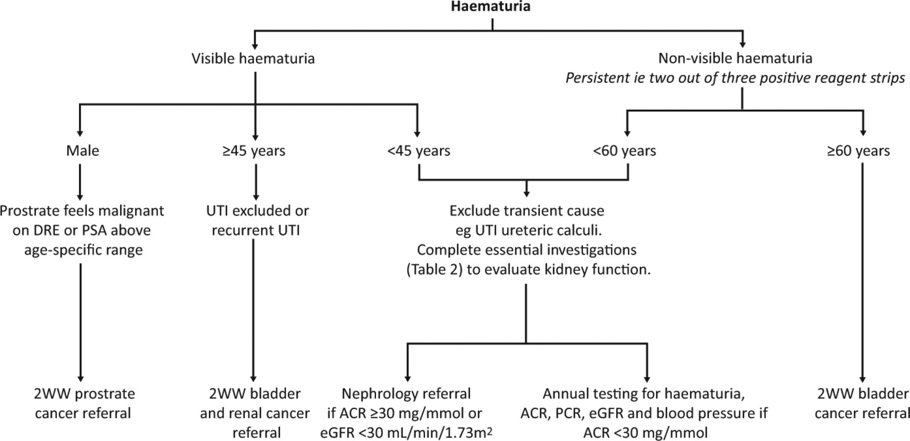
Haematuria
Haematuria may be classified as ‘visible’ or ‘non-visible’. Both can be due to nephrological (eg glomerulonephritis) or urological (eg malignancy, infection or calculi) pathology, and clinical presentation will guide referral. Fig 1 summarises an evaluation process for both visible and non-visible haematuria, adapted from the National Institute for Health and Care Excellence (NICE) guidelines.3,4

Haematuria assessment pathway, adapted from NICE urological cancers clinical knowledge summary and NICE chronic kidney disease guidelines.3,4 ACR = albumin–creatinine ratio; DRE = digital rectal examination; eGFR = estimated glomerular filtration rate; NICE = National Institute for Health and Care Excellence; PCR = protein–creatinine ratio; PSA = prostate specific antigen; 2WW = 2-week wait; UTI = urinary tract infection.
Malignancy
Urological malignancy can cause both visible and non-visible haematuria. History is important in identifying risk factors: smoking status, drug history (eg cyclophosphamide), occupation, chemical exposure and travel history (eg schistosomiasis).5,6 Urgent referral for the assessment of urological malignancy is guided by age (Fig 1). Once malignancy has been excluded, other causes should be considered.
Visible haematuria
A single episode of visible haematuria warrants investigation.7 Transient causes should be excluded by rechecking a urine dipstick after an acute episode has resolved. Anticoagulant and antiplatelet therapy will worsen any haematuria but will not be the precipitant.
The appearance of visible haematuria is a useful indicator to the origin of pathology. Pink stained urine, or frank blood, suggests fresh bleeding along the urinary tract and points towards urological causes. In contrast, visible haematuria that is dark (‘cola-coloured’) is suggestive of a nephrological cause due to haemoglobin being converted to methaemoglobin in the acidic environment.
Co-existent symptoms also provide information. In urological pathology, flank pain suggests ureteric colic, while intercurrent illness (typically upper respiratory tract infection) followed by cola-coloured urine is suggestive of a nephrological cause (eg post-infectious glomerulonephritis or immunoglobulin (Ig) A nephropathy).
Isolated non-visible haematuria with normal renal function
Kidney biopsy to investigate isolated non-visible haematuria, in the absence of proteinuria and renal impairment, with normotension, is unlikely to change management.8,9 Thin-basement membrane nephropathy and IgA nephropathy are frequent causes.8,10 However, persistent isolated non-visible haematuria has been associated with a significant, albeit small, incidence of end-stage kidney disease (ESKD).11,12 Annual monitoring in primary care with blood pressure, urinalysis and serum creatinine levels is necessary.
Biopsy can assist in the diagnosis of inherited conditions and prompt screening of relatives, genetics referral and monitoring. Thin-basement membrane nephropathy usually has a good prognosis with only a minority progressing to ESKD, and so clear diagnosis can provide reassurance and avoid unnecessary further investigation.12 Alport’s syndrome has a lower prevalence but greater risk of progression to ESKD, especially in males, and has extra-renal manifestations, such as hearing impairment.12 Early diagnosis and family screening to identify affected family is essential.
Non-visible haematuria with markers of abnormal renal function
Non-visible haematuria with markers of abnormal kidney function (such as increasing serum creatinine, reduced urine output, hypertension or proteinuria) has a variety of potential urological and nephrological causes.
Regarding intrinsic kidney disease, non-visible haematuria with deranged kidney function often reflects glomerular inflammation (glomerulonephritis). These patients may be systemically unwell and, in the presence of an acute decline in their kidney function, will require close monitoring and follow-up. Kidney biopsy is a key component to diagnosis, management and prognosis, whereby the focus of treatment is the underlying glomerulonephritis, supportive care to manage to complications (eg fluid overload) and to preserve kidney function (eg blood pressure management).13 Biopsy may be postponed or not completed if unlikely to change management, if the patient is high risk or to avoid treatment delays if the diagnosis is clear. For example, in anti-glomerular basement membrane disease, the presence of circulating antibodies, rapidly progressive AKI and haematuria (with or without pulmonary haemorrhage) confirms the diagnosis.14
Proteinuria
Proteinuria should be quantified using both the urinary protein – creatinine ratio (PCR) and albumin–creatinine ratio (ACR). The presence of albuminuria, as defined by an ACR ≥3 mg/mmol, is preferred in the detection and management of chronic kidney disease (CKD) due to its prognostic value and reflection of glomerular injury. Even with an estimated glomerular filtration rate (eGFR) within expected limits, the presence of albuminuria for >3 months reflects persistent glomerular dysfunction and CKD.15 Albuminuria can be subdivided into microalbuminuria (a moderate increase in ACR of ≥3 to ≤30 mg/mmol) and macroalbuminuria (a severe increase in ACR of >30 mg/mmol) influencing decisions regarding monitoring and referral.15 Urinary ACR is an essential investigation when assessing new kidney disease, known CKD and those at risk of kidney damage. This is because urinary ACR will detect early, low-grade proteinuria that has a high risk of being missed on urine dipstick.4
It is important to note that proteinuria can occur as the result of excess serum free light chains or impaired absorption in the proximal tubule. This results in proteinuria with minimal albuminuria (see the myeloma section later), highlighting the risk of false negative results if urine ACR is used in isolation.16
Unless there is a clear history of kidney disease secondary to systemic illness (eg diabetes mellitus and hypertension) or the risks of biopsy outweigh the benefits, biopsy in patients with unexplained proteinuria provides valuable information. The presence of proteinuria should be confirmed on an early morning urine sample. Benign phenomena (such as orthostatic (ie postural) proteinuria) typically occur in individuals <30 years old and presents as isolated proteinuria; the absence of proteinuria on an early morning sample confirms the diagnosis and further investigation is not indicated.17,18
Nephrotic range proteinuria is defined as a urinary PCR >300 mg/mmol or ACR >220 mg/mmol, and may or may not occur with the nephrotic syndrome: heavy proteinuria in combination with hypoalbuminaemia (<25 g/L) and oedema, with or without significant hypercholesterolaemia (>10 mmol/L).15 Nephrotic syndrome is a clinical presentation and the underlying diagnosis should be sought. There are some situations associated with nephrotic syndrome (such as in diabetic kidney disease or amyloidosis) where clinical features and less invasive investigations can indicate the diagnosis and biopsy can be avoided. However, if common clinical diagnoses have been ruled out, it would be important to consider biopsy in adults.
Kidney biopsy in chronic kidney disease vs acute kidney injury
Kidney biopsy is a useful diagnostic tool in both the acute and chronic setting, especially in the presence of active urinary sediment. However, active urinary sediment does not confirm intrinsic kidney disease and biopsy poses unnecessary risk in cases of pre-renal and post-renal causes of kidney impairment. Furthermore, an active urine sediment is not a prerequisite for additional investigation; kidney biopsy may also be appropriate in cases of bland urinary sediment, for instance, non-recovering AKI and suspected tubulointerstitial nephritis.
Specific situations when kidney biopsies may be considered
Diabetic kidney disease
Thirty per cent to 40% of people with type 1 and type 2 diabetes mellitus (DM) in the UK develop CKD and their risk of requiring renal replacement therapy is over three times the general population.19,20 ‘Diabetic kidney disease’ refers to the structural and functional changes caused by DM, while ‘diabetic nephropathy’ refers to specific histological findings on biopsy. Alternative or superadded diagnoses may co-exist (eg hypertensive disease, unresolved AKI or glomerulonephritis).21 The distinction is important for treatment, prognosis and future transplant decisions.
The natural history of diabetic kidney disease in type 1 and type 2 DM is well defined and the development of albuminuria is a consistent predictor for progression to ESKD.22,23 In most patients, clinical history, course of disease and non-invasive investigations can identify where CKD is likely a consequence of DM. National Kidney Foundation guidelines advise that CKD can be attributed to DM in the presence of macroalbuminuria or microalbuminuria with retinopathy, or the presence of microalbuminuria in patients with type 1 DM for >10 years.24 In these instances, the risks of biopsy are thought to outweigh the benefits of a confirmatory diagnosis. Features suggestive of non-diabetic causes of kidney damage that should prompt further investigation (Table 1) include:
refractory hypertension or large drop in renal function following initiation of renin–angiotensin–aldosterone system inhibitors (suggestive of reno-vascular disease)
the absence of diabetic retinopathy in the presence of proteinuria is predictive for non-diabetic kidney disease and strengthens the need for further investigation
haematuria
evidence of other systemic disease.24,25
Additionally, rapidly decreasing kidney function and rapidly increasing proteinuria / nephrotic-range proteinuria may occur due to DM or could indicate an additional diagnoses; further investigation (Table 1) and referral to nephrology would be warranted.
Lupus nephritis
Lupus nephritis (LN) occurs in approximately 50% of patients with systemic lupus erythematosus (SLE).26 Patients may lack overt clinical signs of kidney disease and monitoring kidney function and urinary sediment are important.27 The gold standard for diagnosis of LN is kidney biopsy and earlier biopsy is associated with improved outcomes.28 The role for repeat biopsy in the disease course is also relatively clear.
Initial presentation
LN should be considered in any patient with SLE with declining renal function, proteinuria >0.5 g per 24 hours or active urinary sediment.29,30 Each histological class is associated with different treatment decisions and prognosis; thus, timely biopsy and re-biopsy are essential investigations for both diagnosis and to guide management.31 Biopsy also serves to detect alternative causes of kidney damage in SLE (such as drug-induced nephrotoxicity, lupus podocytopathy or thrombotic microangiopathy).
Repeat biopsy
Biopsy can be used to diagnose relapses or progression of disease. Histological transformation can occur with relapses, potentially changing the treatment required and prognosis.30,31 Furthermore, relapses are an independent predictor of progression to ESKD.32 In patients where relapse is suspected, there is a low threshold for repeat biopsy.29 There are no accurate clinical predictors of class transformation, reinforcing the value in histologically restaging the disease to guide immunosuppression and inform the risk of progression to ESKD.33,34
The older patient
The average age of someone hospitalised with AKI is 76 years.35 Approximately 54% of people aged >75 years live with CKD 3–5, with clear comorbid consequences.36
GFR declines physiologically with age.37 Identifying patients with pathological, non-senescent kidney disease remains challenging. The presence of abnormal urinary sediment warrants investigation, as outlined earlier.
Histological findings may highlight a discrepancy in the clinical diagnosis in up to a third of older patients.38 In a recent UK retrospective cohort study of biopsies in patients aged >70 years, scarring due to diabetes and hypertension was identified in 36% while the remainder included pauci-immune glomerulonephritis (12%), tubulointerstitial nephritis (11%), membranous glomerulonephritis (7%) and other diagnoses.39 Appropriate treatment with immunosuppression in this group can still alter progression to ESKD and survival.40 Biopsies also provide prognostic information; those diagnosed with vasculitis and paraprotein-related renal disease had the highest risk of progression to ESKD compared with other diagnoses.39 Prognostic information facilitates earlier discussions around conservative care or initiation of dialysis. Some studies have found that older patients have an increased risk of bleeding post-biopsy, while others have observed no increased risk compared with other age groups.41,42
As with all data from registries, case series or retrospective cohorts, there will be an element of selection bias involving the population under investigation. However, the findings from these studies highlight that if these patients fit the criteria for requiring a kidney biopsy, then it is generally safe to proceed and age itself should not be an exclusion criterion.
Myeloma
Multiple myeloma is a malignancy of bone marrow plasma cells and is characterised by the clonal proliferation of plasma cells (derived from B cells) and subsequent production of a monoclonal paraprotein.43 Diagnosis involves the presence of end-organ damage attributable to the plasma cell proliferation: hypercalcaemia, bone lesions, anaemia or renal insufficiency.44,45 Up to a third of individuals have kidney impairment at the time of myeloma diagnosis.46 Kidney injury can occur directly (eg light-chain cast nephropathy, immunoglobulin deposition disease or amyloidosis) or indirectly due to sepsis, dehydration, hypercalcaemia or medication toxicity.44,47,48 There may be pre-existing CKD secondary to conditions such as hypertension or DM.45,48 Light-chain cast nephropathy is considered a ‘myeloma-defining’ event.45 Urinalysis will demonstrate proteinuria due to the presence of filtered light chains (Bence–Jones protein), with minimal albuminuria as the glomerular basement membrane is intact.48,49 Histological diagnosis confirms the presence of light-chain cast nephropathy, however, a presumptive diagnosis can be made with high serum free light chain levels (>1,500 mg/L) and AKI; a kidney biopsy should not delay treatment in such cases.45 Biopsy should be considered when a diagnosis is uncertain (for instance, when serum free light chain levels are <500 mg/L) to exclude other causes of AKI and CKD.45,47
Plasma cell dyscrasias in the absence of end-organ damage are termed monoclonal gammopathy of undetermined significance (MGUS) and considered pre-malignant conditions.45 MGUS can lead to kidney injury through glomerular and tubule dysfunction. The term ‘monoclonal gammopathy of renal significance’ (MGRS) has been coined to discriminate non-myeloma plasma cell dyscrasias that result in kidney injury.50 The prognostic significance of biopsy findings in plasma cell dyscrasias is unclear.51 Ultimately, management should be focused on the underlying plasma cell dyscrasia; achieving haematological response is associated with both overall and kidney-specific survival in multiple myeloma and AL amyloidosis.49,52,53
Safety of the procedure
Kidney biopsies are invasive procedures and not without risk. The decision to proceed requires oversight (and usually completion) from the nephrology team and a shared decision with the patient. Biopsies should only be performed when the results will guide treatment, assist with diagnosis that will alter treatment or inform prognosis. Meticulous preparation of the patient is key and important contraindications to biopsy are outlined in Table 2. Ultrasound is important prior to biopsy to ensure that the kidneys can be visualised, to rule out anatomical abnormalities and to provide further information to determine the risk–benefit balance of the procedure. For instance, small kidneys and poor corticomedullary differentiation indicate (unquantifiable) chronicity of the renal disease and potential for limited reversibility, and there may be challenges in differentiating the kidneys from surrounding retroperitoneal structures. Patients may be concerned about the effect of removing kidney tissue on kidney function. Reassuringly, one study estimated that, in stable transplant patients, the GFR loss due to biopsy is 0.77 mL/min.54
Relative and absolute contraindications for kidney biopsy
Major complications from kidney biopsy are related to bleeding: haematoma formation (11%); bleeding requiring transfusion (1.6%); pain (4.3%); macroscopic haematuria (3.5%); and, rarely, death (0.06%).55 Candidates with hypertension, high creatinine, thrombocytopenia, anaemia or requiring early recommencement of anticoagulation are at higher risk of severe bleeding; if these cannot be corrected pre-biopsy and biopsy is essential, then close monitoring is recommended with additional risks discussed with the patient.56–58 Patients should be monitored for 6–8 hours post-procedure and higher risk candidates may be admitted overnight. Any haematuria post-biopsy warrants admission to gain intravenous access and assess full blood count. Computed tomography angiography may be required to identify active bleeding points amenable to endovascular intervention.
It is important to note that histological samples do not always provide a definitive answer and the findings should be interpreted in the context of the history and other investigations. Accurate diagnosis and prognostication can be affected by sampling error, especially in focal pathologies, or where too few glomeruli have been captured. Histological findings are not always specific; for example, interstitial fibrosis and tubular atrophy are signs of chronic damage, rather than pointing towards a diagnosis.
Conclusion
Kidney biopsy is key tool in the evaluation of both AKI, CKD and established intrinsic kidney disease to guide management strategies and confirm diagnoses. It is a procedure not without risk, and so it is essential that preliminary investigations are completed to guide differential diagnoses and secondary care referral, with the exclusion of pre-renal and post-renal causes prior to biopsy completion. Although important, the kidney biopsy should not delay treatment when the diagnosis is established from other investigations.
Key points
Kidney biopsy can be a valuable investigation for acute kidney injury and chronic kidney disease for the diagnosis of intrinsic renal disease and to guide prognosis and management.
Kidney biopsy is an invasive procedure that carries serious, albeit low probability, risks and there are a number of contraindications to consider and should always be undertaken with specialist nephrology input.
Simple investigations, such as serum creatinine, blood pressure, ultrasound of the urinary tract, urine samples (dipstick, PCR, ACR and culture) and a renal immunological screen will assist in guiding whether kidney biopsy is indicated.
Often it is not appropriate to delay treatment to allow for a kidney biopsy if the underlying diagnosis is clear.
A patient-centred approach should be adopted when considering whether a kidney biopsy is appropriate (eg isolated non-visible haematuria or the older patient).
- © Royal College of Physicians 2022. All rights reserved.


