
ABSTRACT
Cardio-oncology is a subspecialty that provides cardiac care for patients with cancer. Newer oncological agents have not only increased survivorship, but also sprouted novel cardiovascular toxicity (CVT) involving any component of the cardiovascular system, albeit with some preferential targets. Patients with cancer should undergo a baseline cardiovascular risk assessment and have individualised surveillance planned during cancer therapy and post treatment. The early diagnosis of CVT, by clinical history and examination along with imaging and laboratory analysis, is paramount. Management includes cardioprotective strategies and multidisciplinary decision-making regarding the risk–benefit ratio of oncological treatment based on CVT.
Key points
Cardio-oncology is an emerging discipline that entails a clinical spectrum of cardiovascular problems that can be encountered either during, immediately after or long after the treatment of cancer.
CVT can involve any structural component of the cardiovascular system but therapeutic agents might have a predominant target.
The goals are to detect and manage the consequences of CVT and consider cardioprotective strategies to mitigate damage and to avoid withholding of cancer therapy.
Long-term follow-up is often required because CVT can appear years later, as seen with chest radiation therapy.
Introduction
Over the past decade, with the appearance of innovative and more effective cancer treatments, survivorship has increased. Most oncological disease occurs in patients in the same age group as patients with cardiovascular disease (CVD) because such patients share similar risk factor profiles. Cardio-oncology is a discipline in which care for patients with either pre-existing CVD or those who develop cardiovascular toxicity (CVT) is provided by a multidisciplinary team comprising cardiologists, oncologists, haematologists, trainees, specialist nurses, physiologists and pharmacists supported by GPs. Several guidelines and consensus statements from various international societies are available to counsel practitioners.1,2 This article addresses the essentials of the discipline relevant to general physicians.
Cardiovascular toxicity related to cancer treatment
Although the classic definition of cardiotoxicity has centred around left ventricular (LV) systolic dysfunction, recent descriptions highlight direct cardiac injury to any of the components of the cardiovascular system, with the potential to cause cardiomyopathy and heart failure, myocarditis, vascular toxicities, arrhythmias, coronary artery disease, premature valve disorders, hypertension and thromboembolism.3 An indirect toxicity can develop through effects on the thyroid, adrenals, pituitary, pancreas and pulmonary vasculature, with downstream consequences on the cardiovascular system.
The three different clinical scenarios include acute CVT, defined while receiving anticancer treatment (eg immune checkpoint inhibitors (ICI) myocarditis); subacute toxicity, during the first 12 months after completion of cardiotoxic treatments (eg LV systolic dysfunction related to HER-2 inhibitors); and long-term, beyond 12 months – cardiovascular complications of previous oncological treatments (eg LV dysfunction related to anthracyclines or constrictive pericarditis following radiation to the chest).
CVT in a patient receiving oncological therapy can be related to chemotherapy, targeted agents, immune therapies, radiation therapy or any combination of agents. Given that novel agents appear in each therapeutic class, CVT specific to each agent is still being recognised and, therefore, assigning class effects should be avoided. Drug interactions between cancer therapies and cardiovascular agents should also be considered. The most frequent causes of cancer treatment-related CVT are summarised in Fig 1.
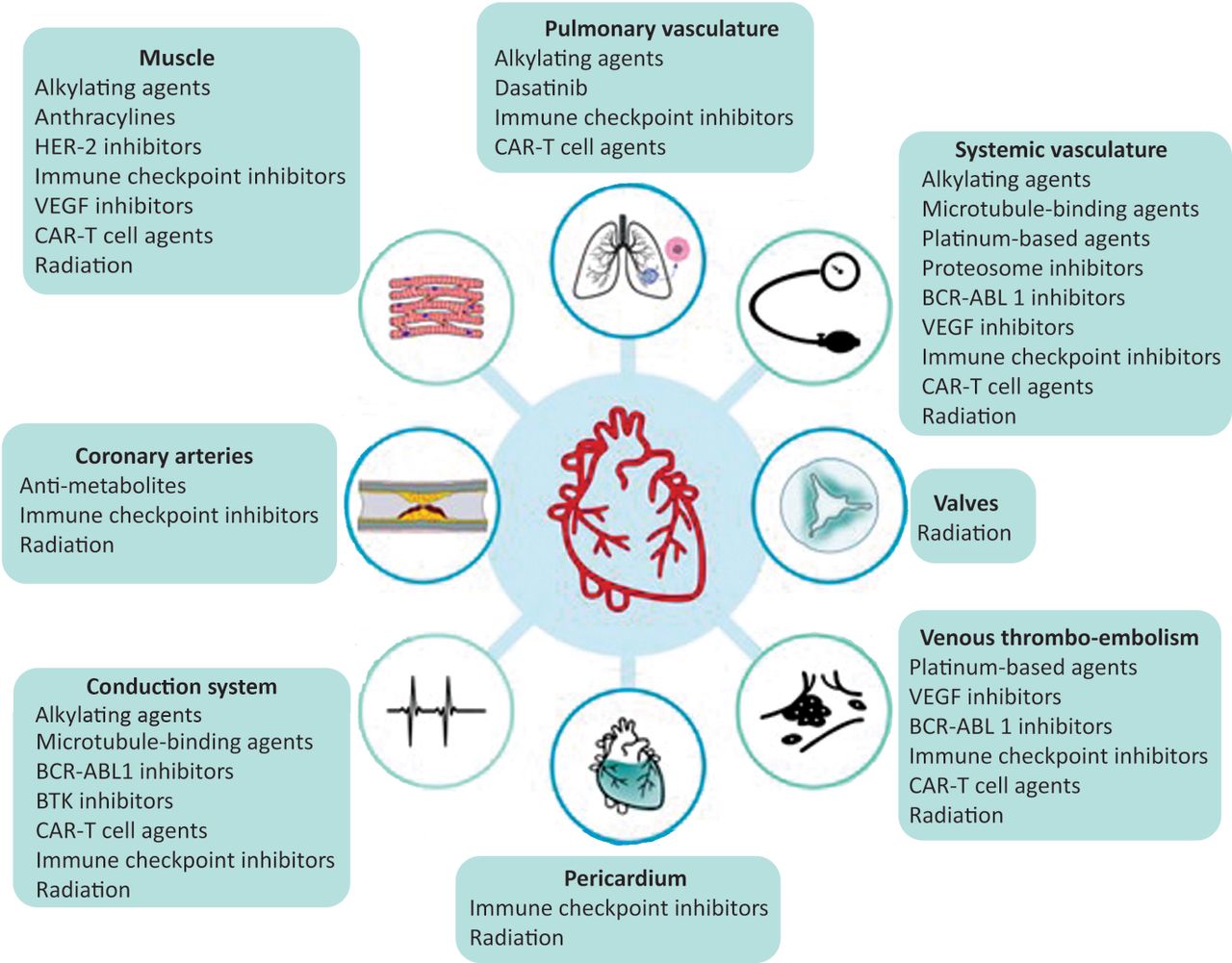
Cancer treatment-related cardiovascular toxicity (CVT). Despite each therapeutic class having the potential to cause multiple CVTs, some oncological drugs have a predilection for certain components of the cardiovascular system. The most common conventional chemotherapies are alkylating agents (eg cyclophosphamide), anthracyclines (eg doxorubicin and epirubicin), antimetabolites (eg 5-fluorouracil (5-FU) and capecitabine), microtubule-binding agents (eg paclitaxel and docetaxel) and platinum-based agents (eg cisplatin). The target therapies more frequently used are human epidermal growth factor receptor 2 (HER-2) inhibitors (eg trastuzumab and pertuzumab), proteosome inhibitors (eg bortezomib and carfilzomib), vascular endothelial growth factor (VEGF) inhibitors (eg bevacizumab and sunitinib), BCR-ABL1 inhibitors (eg dasatinib, nilotinib and ponatinib) and Bruton tyrosine kinase (BTK) inhibitors (e.g. ibrutinib and acalabrutinib). More recent immunotherapies include immune checkpoint inhibitors (eg pembrolizumab, nivolumab and atezolizumab) and chimeric antigen receptor (CAR) T cell therapy. Side effects of radiation usually occur years or even decades following exposure and can affect any cardiovascular structure.
Cardio-oncology patient clinical trajectory
Baseline cardiovascular risk stratification assessment
Baseline clinical assessment for cardiovascular risk factors, history of antecedent CVD and cardiotoxic therapies, physical examination, electrocardiogram and laboratory analysis are mandatory (Fig 2). Although echocardiography is the preferred baseline imaging modality, cardiac magnetic resonance (CMR) and cardiac computed tomography (CT) might also be required.

Cardio-oncology patient clinical trajectory. The patient at the centre is followed up by the multidisciplinary team after diagnosis and during cancer therapy. There has been a paradigm shift in cancer as a chronic disease, because some cancers have sequelae even when they are considered cured. Pretreatment cardiovascular assessment, detection and management of CVT and surveillance of survivors at higher risk for cardiovascular disease (CVD) are key roles of a cardio-oncologist. CAD = coronary artery disease; ECG = electrocardiogram.
Several risk stratification tools have been designed to grade patients into low, moderate, high or very high risk of CVT before starting treatment.4 Some guidelines have incorporated the Heart Failure Association/International Cardio-Oncology Society risk assessment tool.5 In clinical practice, even though these calculators can be useful, it is vital that clinicians adopt a systematic approach avoiding the tendency to delay oncological treatment, because therapy for cancer is time sensitive and any delay can adversely affect outcomes.
A personalised cardiac surveillance and prevention strategy, based on baseline CVT risk, type and stage of cancer and proposed oncological therapy, is formulated at first review with a cardio-oncologist. Prevention strategies include measures to optimise lifestyle choices and mitigate traditional cardiovascular risk factors. In those with high or very high risk of CVT, guideline-directed cardioprotective heart failure (HF) therapy or dexrazoxane/liposomal anthracyclines, for those treated with anthracyclines, should be considered.
Diagnosis and monitoring of CVT
Regular clinical evaluation and physical examination are recommended during cancer treatment to detect early symptoms and signs of CVT. Electrocardiograms are required in patients at risk of cardiac arrhythmias or QTc prolongation according to specific drug protocols. Cardiac serum biomarkers, such as natriuretic peptides and cardiac troponin, can be helpful to detect and monitor CVT or to attribute a cardiac cause to unspecified symptoms, such as shortness of breath.
Echocardiography can detect deterioration of LV function by measurement of ejection fraction with conventional techniques or more refined ones, such as 3D echocardiography. Assessment of global longitudinal strain, a marker of early LV subclinical dysfunction, provides additional information.4 CMR in patients with poor image quality or when the echocardiogram is not diagnostic can detect both functional and structural changes assessing cardiac function, cardiac invasion and ischaemia. CMR is particularly helpful for the tissue characterisation of cardiac masses and the diagnosis of myocarditis. However, CMR is limited by its availability and cost.6
Cardiac CT can be used to assess the coronary arteries to exclude acute coronary syndromes and assess cardiac masses. In some cases, invasive coronary angiography might be required to directly visualise the coronary arteries. Nuclear imaging can also have a role in specific situations, such as cardiac amyloidosis and interrogation of metabolic activity of suspicious cardiac masses.
Management of CVT
Cancer treatment-related cardiac dysfunction
The cancer treatment-related cardiac dysfunction (CTRCD) that typically occurs with anthracycline and HER-2 inhibitors might present clinically or be detected in asymptomatic patients during surveillance. Temporary interruption of treatment is recommended in patients who develop moderate to severe CTRCD and rechallenge can be considered, using cardioprotective strategies with close monitoring.7 Cardioprotective strategies include minimising the drug dose and, in the case of anthracycline, switching to liposomal anthracycline preparations or pretreatment with dexrazoxane before each further cycle. Guideline-directed HF therapy is recommended in patients who develop symptomatic CVT or moderate to severe CTRCD. The use of an angiotensin-converting enzyme inhibitor/angiotensin II receptor blocker (ACEi/ARB) or angiotensin receptor–neprilysin inhibitor (ARNI), a beta-blocker, a mineralocorticoid receptor antagonist (MRA), and a sodium–glucose co-transporter 2 inhibitor (SGLT2i) is recommended, with uptitration to target doses as tolerated. Notably, most clinical trials in HF excluded patients with cancer, reflecting the use of these agents in an extrapolated manner. Frequent cardiac surveillance with imaging and serum biomarkers is recommended in all patients with CTRCD.
Myocarditis
Myocarditis is a rare but severe complication of ICI, characterised by severe cardiovascular symptoms, a new increase in troponin and electrocardiographic abnormalities (atrioventricular or ventricular conduction disorders, bradycardia and tachyarrhythmias). Interruption of ICI is recommended and treatment with high-dose methylprednisolone should be promptly initiated in patients who are haemodynamically unstable, while awaiting further confirmatory testing with echocardiogram and CMR.
Acute coronary syndrome
Diagnosis of acute coronary syndrome (ACS) is based on the same principles as in patients without cancer, including symptoms, an early 12-lead electrocardiogram, and serial measurements of troponin. Cancer treatment should be temporarily interrupted, and an individualised action plan, considering the severity of ACS, the cancer status and prognosis, is prudent. Some agents, such as fluoropyrimidines, can induce coronary vasospasm and myocardial ischaemia at rest several days later after their administration.
Arrhythmias
Chemotherapy, radiotherapy, cancer surgery, cancer itself (invasion), related electrolyte disturbances or pre-existing substrates can increase the risk of arrhythmias. Atrial fibrillation is the most prevalent arrhythmia and the risk–benefit ratio of long-term anticoagulation in patients with cancer is a major concern. It is important to realise that the CHA2DS2-VASC score is not validated specifically in cardio-oncology patients. Cancer therapy-induced ventricular arrhythmias are rare and are related to a prolongation of QTc leading to the development of torsade de pointes. Immediate withdrawal of any QT prolongation drug, correction of electrolyte abnormalities and close electrocardiographic monitoring are essential.
Arterial hypertension
Arterial hypertension in patients with cancer can be caused by their oncological treatments, non-cancer drugs and other factors, including stress, pain, renal impairment or metabolic factors.8 Given their well-established cardioprotective effects, ACEi or ARB are first-line therapies in this context.
Radiation induced-coronary artery, valve and pericardial disease
Many years after radiation therapy, damage can be detected in the coronary arteries, cardiac valves and the pericardium, especially if the cardiac chambers are in the field of therapy. Dose modulation and adequate chest shielding should be used.
Cardiac masses and infiltration
Both CMR and CT can help differentiate between tumour and thrombus in the cardiac chambers. With its ability to identify metabolic activity, positron emission tomography (PET) can help identify primary and secondary tumours.
Follow-up of long-term survivors after therapy: late effects
In the first year after treatment, risk assessment for CVT should be done to establish a long-term follow-up plan. The development and severity of CVT is influenced by several variables, including the types and doses of cumulative treatment received, the time elapsed since treatment, and pre-existing CVD. Therefore, a long-term survivor surveillance strategy is required to identify those at high risk of developing late effects, particularly patients who have received high-dose doxorubicin and prior radiation.9 Cardiac rehabilitation for those who have functional deterioration is also important.
Future requirements in cardio-oncology
Cardio-oncology services and care networks are essential to integrate services.
An established structure of training in cardio-oncology.
Improved risk prediction and diagnostic tools and their integration in a synergistic manner to better improve prognosis.
Clinical trials to specifically address cancer treatment-related CVT.
Conclusions
Cardio-oncology has grown into a new subspecialty in the UK over the past decade, with expansion of several comprehensive and multidisciplinary cardio-oncology units. Value provided by cardio-oncologists includes developing tailored management strategies for patients with cancer despite the limited, albeit growing, evidence base. Continued interest from oncologists and cardiologists across the UK is required to establish new services and for current ones to develop further. Specialist cardio-oncologists alone cannot care for the number of survivors of cancer with cardiovascular issues, and knowledge sharing should be a priority.10 To improve both cardiovascular and cancer outcomes, GPs and general physicians should be cognisant of the wide variety of potential cardiovascular issues in oncological patients. Ultimately, if all hospitals have local cardio-oncology ‘champions’ (ie a cardiologist and an oncologist with a special interest in cardio-oncology) this will be a big step forward in the management of CVT in patients with cancer.
- © Royal College of Physicians 2023. All rights reserved.


