
Main menu

  • Home
  • Our journals
    • Clinical Medicine
    • Future Healthcare Journal
  • Subject collections
  • About the RCP
  • Contact us

Clinical Medicine Journal

  • ClinMed Home
  • Content
    • Current
    • Ahead of print
    • Archive
  • Author guidance
    • Instructions for authors
    • Submit online
  • About ClinMed
    • Scope
    • Editorial board
    • Policies
    • Information for reviewers
    • Advertising

User menu

  • Log in

Search

  • Advanced search
RCP Journals
Home
  • Log in
  • Home
  • Our journals
    • Clinical Medicine
    • Future Healthcare Journal
  • Subject collections
  • About the RCP
  • Contact us
Advanced

Clinical Medicine Journal

clinmedicine Logo
  • ClinMed Home
  • Content
    • Current
    • Ahead of print
    • Archive
  • Author guidance
    • Instructions for authors
    • Submit online
  • About ClinMed
    • Scope
    • Editorial board
    • Policies
    • Information for reviewers
    • Advertising

Severe hypercalcaemia mimicking acute myocardial infarction

Laura C Wesson, Venk Suresh and Rob G Parry
Download PDF
DOI: https://doi.org/10.7861/clinmedicine.9-2-186
Clin Med April 2009
Laura C Wesson
Royal Cornwall Hospital
Roles: Specialist Registrar in Geriatrics
  • Find this author on Google Scholar
  • Find this author on PubMed
  • Search for this author on this site
Venk Suresh
Derriford Hospital, Plymouth
Roles: Specialist Registrar in Cardiology
  • Find this author on Google Scholar
  • Find this author on PubMed
  • Search for this author on this site
Rob G Parry
Royal Cornwall Hospital
Roles: Consultant Nephrologist
  • Find this author on Google Scholar
  • Find this author on PubMed
  • Search for this author on this site
  • Article
  • Figures & Data
  • Info & Metrics
Loading

Abstract

Significant hypercalcaemia can cause electrocardiogram (ECG) changes mimicking an acute myocardial infarction. It is important to recognise that some ECG changes are due to conditions other than cardiac disease so that appropriate treatment is given, and importantly, inappropriate treatments are avoided.

Lesson

A 69-year-old woman presented to the accident and emergency department with an electrocardiogram (ECG) showing anterior ST segment elevation. She was found collapsed at home two hours prior to presentation with vague chest pain, lethargy and being non-specifically unwell. Little history was available from the patient due to consciousness level and a weak voice. Past medical history included ischaemic heart disease, previous coronary angioplasty, left ventricular failure, hypertension, hypercholestrolaemia and ongoing investigation of a thyroid goitre.

On examination she looked unwell, dehydrated, frail but haemodynamically stable. She was not sweaty, clammy or in pain. Cardiovascular system examination was unremarkable. Her initial ECG showed widespread ST segment elevation in leads V1 to V5, Q waves and poor R wave progression in inferior leads (Fig 1). She received thrombolysis on the basis of a significant cardiac history, and significant ECG changes associated with collapse.

Fig. 1.
  • Download figure
  • Open in new tab
  • Download powerpoint
Fig. 1.

Electrocardiogram on admission.

Subsequently, total creatinine kinase and troponin T values were found not to be elevated, but her corrected calcium was later found to be elevated at 5.09 mmol/l. A thyroid gland biopsy result, taken two weeks previously, revealed disseminated thyroid cancer. She rapidly deteriorated despite aggressive resuscitation and died within 36 hours of presentation with refractory hypercalcaemia. No evolutionary changes typical for ST elevation myocardial infarction (MI) were seen (Fig 2). Retrospectively, she was profoundly unwell from hypercalcaemia due to a primary thyroid malignancy on presentation, but treatment was distracted by the dramatic and unexplained ECG.

Fig. 2.
  • Download figure
  • Open in new tab
  • Download powerpoint
Fig. 2.

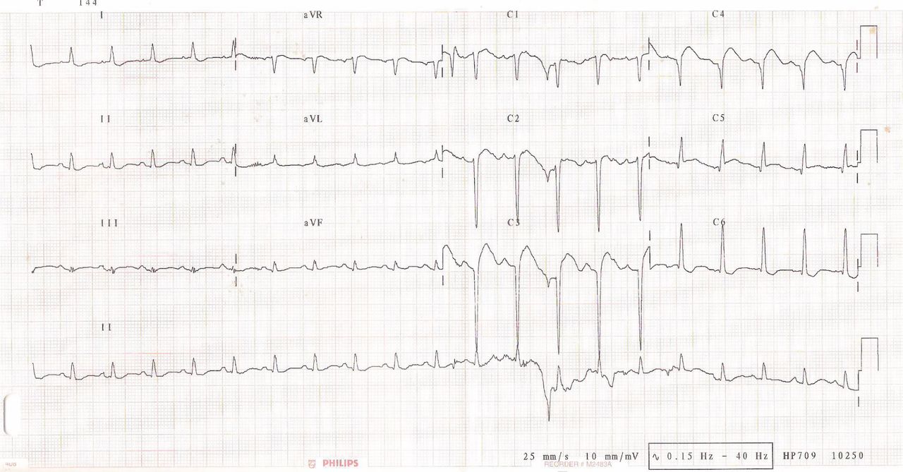
Electrocardiogram pre-terminal.

Discussion

We are aware of only two cases of hypercalcaemia causing ECG changes mimicking acute MI.1,2 The first case was due to hyperparathyroidism, and the second due to vitamin D intoxication. There is also one case, in a series of 161 fatal thyroid carcinoma cases, where the patient died of hypercalcaemia, while another nine cases had hypercalcaemia per se.3 In this series it states that malignancy associated hypercalcaemia can be due to humoral (PTH driven) hypercalcaemia or bone metastases. In this patient hypercalcaemia could be due to either mechanism.

The mechanism of how hypercalcaemia causes ST elevation is not understood, but presumably has a basis in ion concentrations mediating abnormalities in myocardial membrane potentials. To function correctly, individual myocardial cells rely on normal concentrations of biochemical parameters. Classically hypercalcaemia increases myocardial contractility and irratibility. Classical ECG features of hypercalcaemia are absent or shortened ST segment, shortened QT but lengthened T wave duration. It is hoped that this lesson with increase awareness that hypercalcaemia can also cause ECG changes mimicking acute MI. It is important to recognise that some ECG changes are due to conditions other than cardiac disease so that appropriate treatment is given early, and inappropriate thrombolysis avoided.

  • © 2009 Royal College of Physicians

Reference

  1. ↵
    Turhan S, Kilickap M, Kilinc S. ST segment elevation mimicking acute myocardial infarction in hypercalcaemia. Heart 2005; 91:999.doi:10.1136/hrt.2003.030874
    OpenUrlFREE Full Text
  2. ↵
    Ashizawa N, Arakawa S, Koide Y et al. Hypercalaemia due to vitamin D intoxication with clinical features mimicking acute myocardial infarction. Intern Med 2003; 42:340–4.doi:10.2169/internalmedicine.42.340
    OpenUrlCrossRefPubMed
  3. ↵
    Kitamura Y, Shimizu K, Nagahama M et al. Immediate causes of death in thyroid carcinoma: clinicopathological analysis of 161 fatal cases. J Clin Endocrinol Metab 1999; 84:4043–9.doi:10.1210/jc.84.11.4043
    OpenUrlCrossRefPubMed
Back to top
Previous articleNext article

Article Tools

Download PDF
Article Alerts
Sign In to Email Alerts with your Email Address
Citation Tools
LESSON OF THE MONTH (1)
Laura C Wesson, Venk Suresh, Rob G Parry
Clinical Medicine Apr 2009, 9 (2) 186-187; DOI: 10.7861/clinmedicine.9-2-186

Citation Manager Formats

  • BibTeX
  • Bookends
  • EasyBib
  • EndNote (tagged)
  • EndNote 8 (xml)
  • Medlars
  • Mendeley
  • Papers
  • RefWorks Tagged
  • Ref Manager
  • RIS
  • Zotero
Share
LESSON OF THE MONTH (1)
Laura C Wesson, Venk Suresh, Rob G Parry
Clinical Medicine Apr 2009, 9 (2) 186-187; DOI: 10.7861/clinmedicine.9-2-186
del.icio.us logo Digg logo Reddit logo Twitter logo CiteULike logo Facebook logo Google logo Mendeley logo
  • Tweet Widget
  • Facebook Like
  • Google Plus One

Jump to section

  • Article
    • Abstract
    • Lesson
    • Discussion
    • Reference
  • Figures & Data
  • Info & Metrics

Related Articles

  • No related articles found.
  • PubMed
  • Google Scholar

Cited By...

  • Synthetic biology-based cellular biomedical tattoo for detection of hypercalcemia associated with cancer
  • Not all ST-segment changes are myocardial injury: hypercalcaemia-induced ST-segment elevation
  • Google Scholar

More in this TOC Section

  • Lesson of the month 1: Broken heart in the intensive care unit
  • Lesson of the month 1: Sudden onset postural livedo reticularis, cyanotic toes and multiorgan failure
  • Lesson of the month (1): Cabergoline – ‘I eat funny on that’
Show more Lesson of the month (1)

Similar Articles

Navigate this Journal

  • Journal Home
  • Current Issue
  • Ahead of Print
  • Archive

Related Links

  • ClinMed - Home
  • FHJ - Home
clinmedicine Footer Logo
  • Home
  • Journals
  • Contact us
  • Advertise
HighWire Press, Inc.

Follow Us:

  • Follow HighWire Origins on Twitter
  • Visit HighWire Origins on Facebook

Copyright © 2021 by the Royal College of Physicians