
Experience of dermatological problems presenting to a large adult haemato-oncological centre (King's College Hospital, London) indicates 10 main haematological malignancies of interest to dermatologists (Table 1). The skin complications may be conveniently classified as infective, paraneoplastic, autoimmune or infiltrative, or be the result of treatment. The haematological diagnosis is often already established before any skin signs emerge (eg leukaemia cutis), but in some cases skin lesions may be the presenting feature (eg Sweet's syndrome, infection, purpura) or be the sole manifestation with possible subsequent systemic spread (eg cutaneous T-cell lymphoma).
Main haematological malignancies of concern to dermatologists.
Cutaneous manifestations of infection in haematological malignancy
The immunocompromised, and often neutropenic, haematology patient is at risk from various bacterial, viral and fungal infections. Any organism may become pathogenic, including commensals and even environmental organisms such as Saccharomyces cerevisiae (Baker's yeast).1In the presence of severe neutropenia, the physical signs of inflammation associated with infection may be modified or diminished.
Bacterial infection
The skin may be involved primarily or secondarily to disease elsewhere.
Primary infections. Staphylococcus aureus may cause abscesses or paronychia. Methicillin-resistant gram-positive staphylococci pose a particular hazard. Cellulitis (erysipelas) may be caused by S. aureus or more commonly Streptococcus pyogenes. Pseudomonas aeruginosa causes ecthyma gangrenosum where single or multiple ulcerative lesions with a necrotic centre and surrounding induration have a predilection for the perineum, axillae and submammary areas.
Secondary infections. Infections with mycobacterium species (M. kansasii, M. avium intracellulare complex and M. chelonae) are rare but may present with granulomatous papules or nodules in the skin. Nocardiasis should be considered in the presence of cellulitis or erythema nodosum.2
Fungal infection
Primary infections. Mucosal candidosis is readily encountered. Pityriasis versicolor, a superficial yeast infection due to overgrowth of the commensal Pityrosporum orbiculare, is common in the immunosuppressed, especially in patients with lymphoma.
Systemic infections. The most common fungal pathogens which may involve the skin are candida, aspergillus and fusarium, presenting as tender red macules with central purpura. Skin lesions are present in 70% of patients with disseminated fusarium.3Mortality is high in neutropenic hosts with disseminated disease and antifungal therapy alone is often inadequate. The underlying malignancy must be treated.
Viral infection
Reactivation of latent infections such as varicella zoster virus (often resulting in disseminated shingles), human papilloma virus (widespread warts (Fig 1)), herpes simplex virus (‘cold sores’) and mollusca are common. Cytomegalovirus (CMV) only occasionally involves the skin. Patients with lymphoproliferative disorders and chronic lymphocytic leukaemia (CLL) are particularly prone. Prolonged T-cell mediated immunosuppression associated with chemotherapy (especially ciclosporin, tacrolimus and systemic steroids) and haematopoietic stem-cell transplantation facilitates reactivation of viruses.
Cutaneous paraneoplastic and autoimmune associations of haematological malignancy
Neutrophilic dermatoses
Sweet's syndrome and pyoderma gangrenosum are the commonest of the neutrophilic dermatoses. They are cutaneous disorders characterised pathologically by superficial or deep dermal infiltrates of normal appearing neutrophils with no evidence of sepsis. The pathogenesis is unclear.
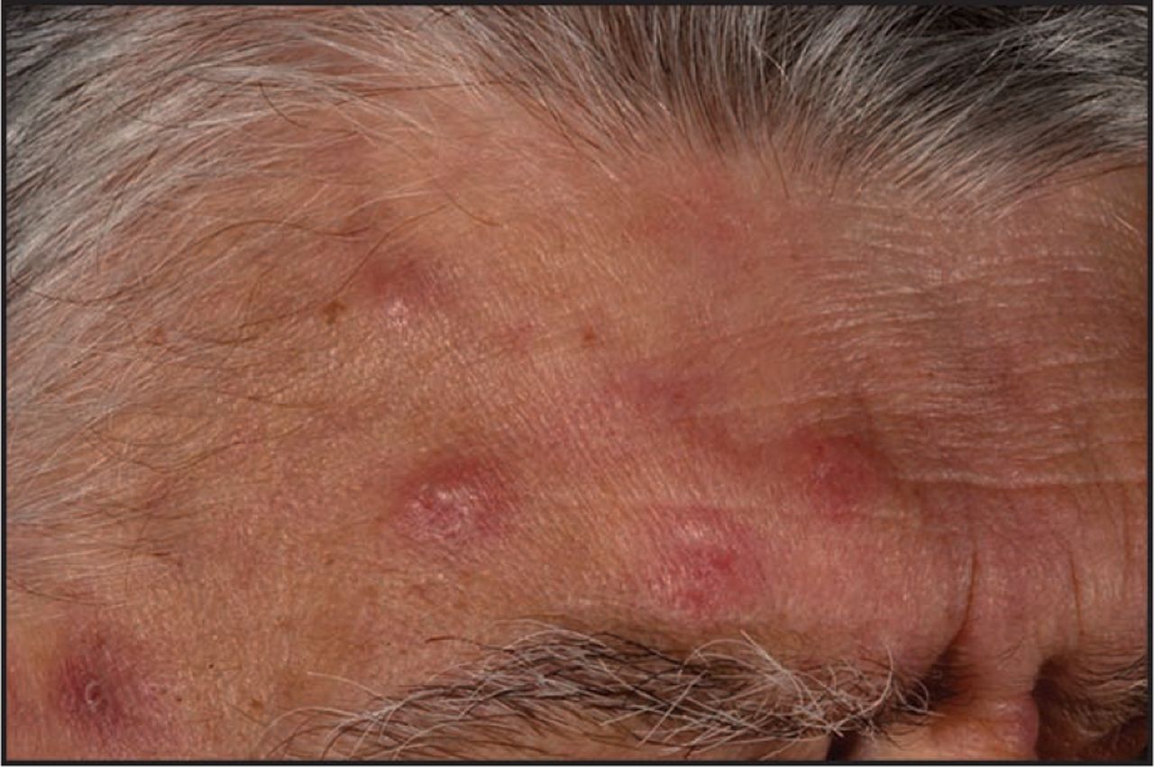
Sweet's syndrome (Fig 2). In this form of neutrophilic dermatosis, the lesions are debilitating, well-defined red or plum coloured plaques or nodules, sometimes with pustules on the surface. They may occur anywhere on the body including the face, associated with fever and neutrophilia. Lesions are frequently misdiagnosed as abscesses and incorrectly treated with antibiotics. The syndrome is responsive to systemic steroids. Often associated with myelodysplastic syndrome (MDS) and acute myeloid leukaemia (AML),4 it may also be induced by granulocyte colony-stimulating-factor (G-CSF) (including those healthy donors who receive G-CSF prior to bone marrow transplantation).5

Disseminated warts. On presenting to a dermatologist this woman was found to be lymphopenic. HIV testing was negative. A bone marrow biopsy performed by a haematologist showed non-Hodgkin's lymphoma. The warts disappeared with chemotherapy.

Sweet's syndrome. This man with myelodysplastic syndrome developed infiltrated, red plaques. Biopsy showed a sterile neutrophilic infiltrate.
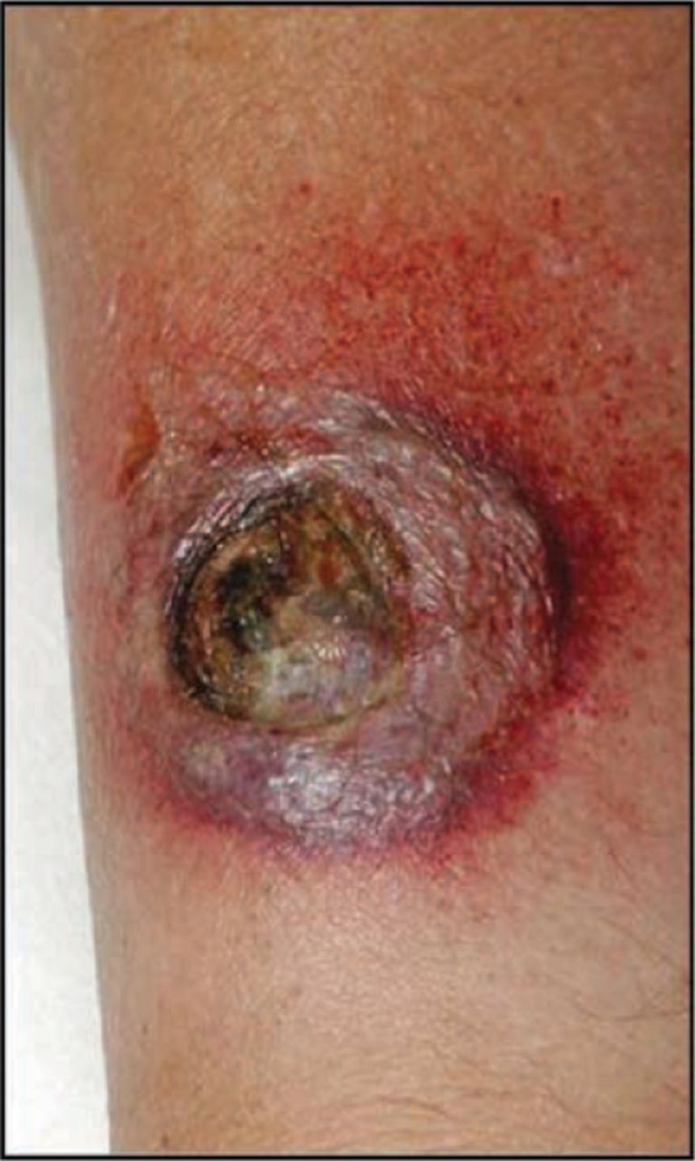
Pyoderma gangrenosum (Fig 3). This condition comprises painful red nodules which rapidly break down to become ulcers with a marked bluish undermined overhanging edge which fail to heal. Lesions frequently develop at the sites of medical intervention (such as Hickman lines and intravenous access sites) and, having been misdiagnosed as sepsis, are often debrided. In 7% of cases pyoderma gangrenosum is associated with haematological malignancies (including AML, chronic myeloid leukaemia and multiple myeloma (MM)), heralding an adverse prognosis and systemic sequelae.6It is also associated with inflammatory bowel disease and rheumatoid arthritis. It responds rapidly to oral steroids and ciclosporin.

Pyoderma gangrenosum. An ‘infection’ developed at the site of the Hickman line and at other venous access sites in this patient. It was debrided until a haematologist made the correct diagnosis and a biopsy was performed. The lesions responded dramatically to steroids.
Autoimmune skin disorders
Thyroiditis, thrombocytopenic purpura and haemolytic anaemia are examples of increased autoimmune phenomena in MDS, possibly due to the complex immune dysfunction present in this disease. Cutaneous autoimmune disorders are also more common, including alopecia areata, vitiligo, cutaneous vasculitis, Raynaud's phenomena, scleroderma and eczema.7In addition, similar autoimmune-related skin conditions are seen following bone marrow transplantation.
Paraneoplastic pemphigus
Paraneoplastic pemphigus is rare. There is usually an intractable painful stomatitis with involvement of the oropharynx and lips. The skin lesions are polymorphic, simulating lichen planus and erythema multiforme, but flaccid blisters which become eroded are usually evident. Three-quarters of cases are associated with lymphoproliferative disorders including non-Hodgkin's lymphoma (NHL), CLL and Castleman's disease (angiofollicular hyperplasia).8
Miscellaneous
Hodgkin's disease (HD), NHL and myelo-proliferative disorders can present with intractable pruritus. Aquagenic urticaria is particularly associated with polycythaemia rubra vera. Acquired ichthyosis is associated with HD, often preceding systemic signs. Diffuse rhomboid scales on the trunk and extensor surfaces are present. Erythema nodosum has also been associated with HD.8
Cutaneous signs of infiltrative disease and malignancy
Leukaemia cutis is an infiltration of neoplastic leukocytes into the skin, presenting as deep red or purple plaques or nodules. It occurs in most haematological malignancies, especially AML, and generally indicates a poor prognosis.9Myeloma may occasionally involve the skin in a similar way.
Adult T-cell leukaemia/lymphoma associated with human T-cell leukaemic virus type-1 retrovirus infection, is characterised by lymphadenopathy, hepatosplenomegaly, hypercalcaemia and central nervous system involvement. The skin is frequently involved early on and mimics the widespread, atrophic, irregular-shaped patches, plaques and nodules of cutaneous T-cell lymphomas such as mycosis fungoides. There are some cases which have only skin signs.10
An increased incidence of primary skin malignancies, which may be clinically and histologically atypical, is reported in patients with CLL.11They may also occur following either radiotherapy for lymphoma or photochemotherapy for the itch associated with polycythaemia rubra vera.
Plasma cell disorders
MM has some specific cutaneous associations. It is classically associated with amyloid deposition in blood vessels, leading to purpura around the eyes and intra-orally, precipitated by coughing or straining to pass a stool. Infiltration of the dermis with amyloid protein causes thickening of the fingers and nails may become dystrophic. Cryoglobulinaemia (type 1) is seen in association with abnormal circulating proteins in MM (and CLL). A diffuse pattern of plane xanthomata, particularly on the face and associated with normolipaemia, is a rare complication, possibly due to paraprotein-lipoprotein complex deposits in the skin.
The syndrome of polyneuropathy, organomegaly, endocrinopathy, monoclonal gammopathy, and skin changes (POEMS) is a rare multisystem disease associated with plasma cell disorders. The common skin changes are hyperpigmentation, skin thickening, scleroderma-like changes, hypertrichosis and haemangiomas which have a histology specific to the syndrome (glomeruloid haemangioma).
Cutaneous manifestations of therapies used in haemato-oncology
Drugs
Toxic erythema (a generalised morbiliform eruption) is the commonest reaction to drugs, often an antibiotic. Vancomycin, widely prescribed in haematology, may cause a morbiliform drug eruption with a specific linear immunoglobulin A (IgA) pattern on immunofluorescence.12
Antimetabolites, such as fludarabine, which interfere with DNA synthesis, and anthracyclines, which are directly toxic DNA/RNA intercalating agents, may cause similar maculopapular rashes and stomatitis. Antifungal agents (eg poza-conazole and voriconazole), prescribed both as treatment and prophylaxis, cause photosensitivity.
Stevens-Johnson syndrome and toxic epidermal necrolysis are much less common but are serious drug hypersensitivity reactions characterised by varying degrees of rapid and painful epidermal shedding with mucosal surface involvement. Drug rash with eosinophilia and systemic symptoms (DRESS) may also be encountered; there is a symmetrical, red exanthematous eruption with lymphadenopathy and variable organ involvement.
As new therapies are developed, more drug hypersensitivity reactions are being described, including widespread cutaneous reactions to tyrosine kinase inhibitors such as imatinib.13An example is characteristic loss of pigmentation around the eyes (‘panda eyes’) (Fig 4). The demethylating agent, 5-azacytidine, used to treat MDS, can cause lipoatrophy on the face. Bortezomib, a proteasome inhibitor used in MM, reportedly causes a small-vessel necrotising vasculitis.
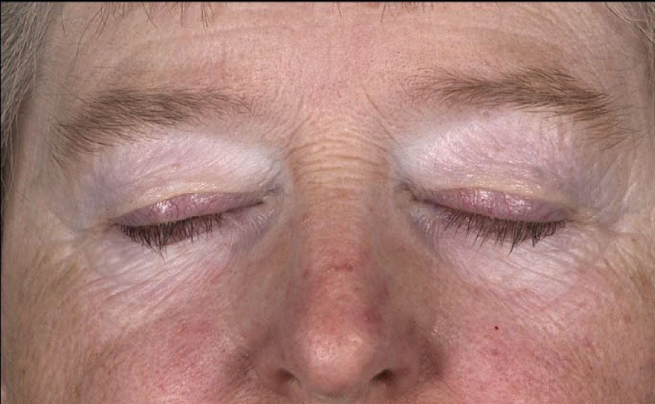
Panda eyes. While taking imatinib for chronic myeloid leukaemia this woman developed depigmentation around her eyes, simulating vitiligo. This is a not an unusual complication.
Key points
The skin is frequently involved in haematological malignancy. A dermatological opinion is critical
The usual physical signs of inflammation associated with infection may be modified in the presence of severe neutropenia
Any organism, be it a skin commensal or ubiquitous environmental organism, may become pathogenic in the immunocompromised neutropenic host
Early skin biopsy can aid accurate diagnosis of skin
More specific eruptions associated with chemotherapy are:
the palmar-plantar erythema dysaesthesia syndrome, and
neutrophilic eccrine hidradenitis (Fig 5) (erythematous multiple or single pur-puric macules, papules or nodules), a rare but distinctive disorder involving sweat glands, which is fairly specific to cytarabine and doxorubicin therapy.
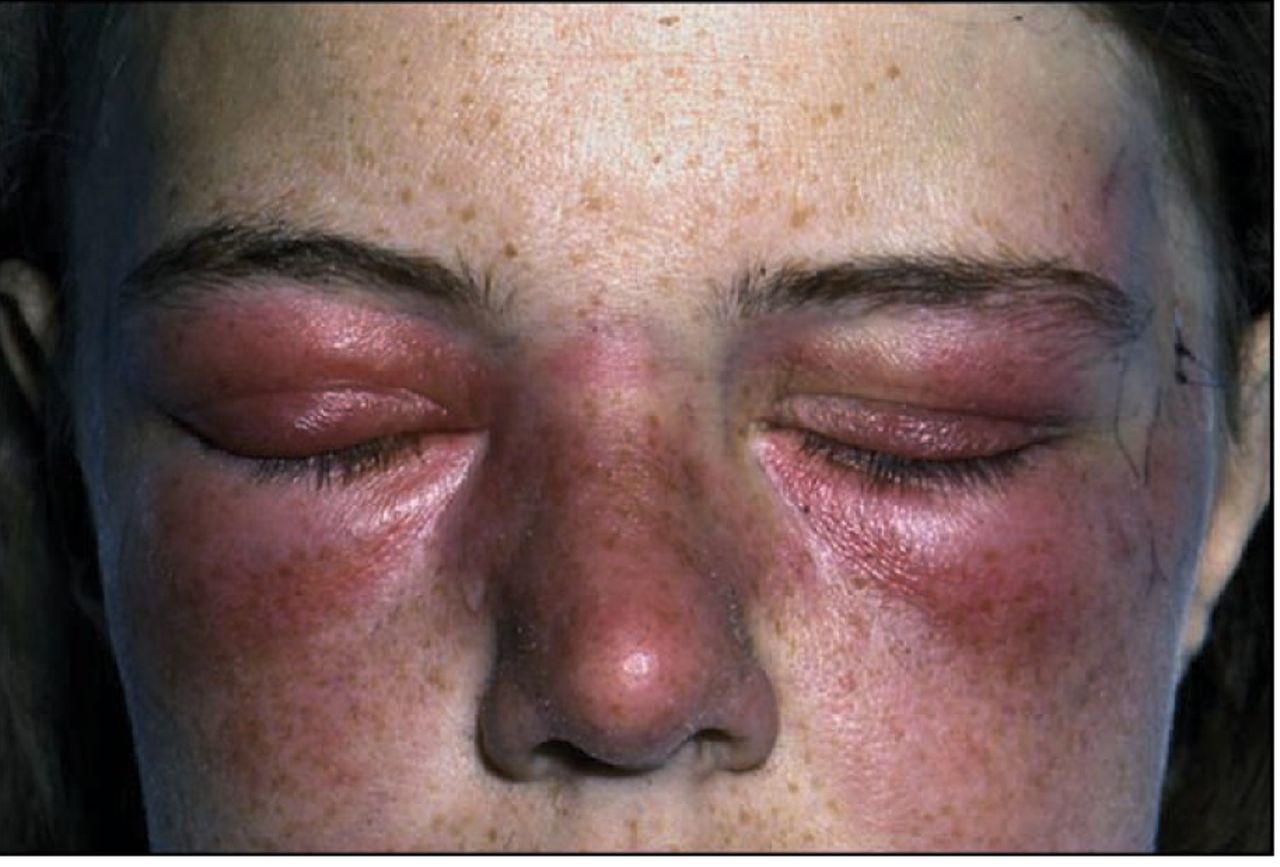
Neutrophilic eccrine hidradenitis. The mauve discoloration and swelling around the eyes is almost diagnostic. A skin biopsy showed a neutrophilic infiltration around the sweat glands. It was due to cytarabine.
Graft-versus-host disease
Graft-versus-host disease (GVHD) occurs in 50% of recipients of allogeneic haematopoietic stem cell transplants14 induced by immune-competent donor cells attacking host tissues. The skin (often affected first), gastrointestinal tract and liver are the commonest targets but other organs can also be affected. Cutaneous GVHD has been classified into acute (100 days post-transplant, typically 14–42 days) and chronic (≥100 days after transplant). Traditionally, acute cutaneous GVHD is considered a widespread, symmetrical, maculopapular or morbiliform exanthema with a predilection for the palms and soles, while chronic GVHD is a spectrum of eruptions simulating lichen planus and connective tissue disorders including scleroderma and lichen sclerosis.15
However, classification in this way may now be outdated. With novel therapies, clinical manifestations of acute GVHD can be seen after 100 days. Conversely, signs of chronic GVHD may be seen relatively early.
A novel form of eczematoid GVHD has recently been described.16This is a distressing eczematous, erythrodermic eruption (Fig 6) with histological features of GVHD. It may present early or late post-transplant but is chronic in its behaviour. It is seen more commonly after either a second transplant or donor lymphocyte infusion and in association with reactivation of CMV.

Chronic eczematous graft-versus-host disease. The patient was erythrodermic (universal involvement of the skin), unwell and shivering from profound loss of fluid, protein and heat from the skin. He was given systemic steroids and tacrolimus but was weaned off them with photochemotherapy (PUVA).
Cutaneous GVHD can be a most disabling condition. Current treatment options are topical corticosteroids or tacrolimus for acute GVHD with less than 50% body surface involvement,17 but oral steroids and ciclosporin or tacrolimus may be necessary. In chronic disease, photochemotherapy (psoralen plus UVA) may be effective, especially for the erythrodermic variety and may reduce the risk of infection and reactivation of the leukaemia associated with immunosuppressive therapy. Extracorporeal photophoresis may be helpful in sclerodermatous GVHD.
Conclusions
Patients with haemato-oncological malignancies are frequently unwell. When a skin eruption is present access to a dermatological opinion is essential for an astute clinical diagnosis. A skin biopsy should be performed, where appropriate, to permit accurate histological, immuno-cytochemical and microbiological analysis. In addition, polymerase chain reaction-based molecular techniques improve the speed and accuracy of diagnosis of infections.
Effective management of the cutaneous manifestations also clearly requires the treatment of the underlying haematological malignancy.
Acknowledgements
We would like to acknowledge the helpful criticism of this manuscript by Dr Stephen Devereux, Consultant Haematologist and Honorary Senior Lecturer, King's College Hospital, London, and the privilege of working with Professor Ghulam Mufti and his haematological colleagues (Dr Steven Schey, Dr Aloysius Ho, Dr Antonio Pagliuca and Dr Robert Marcus) at King's College Hospital.
- © 2009 Royal College of Physicians
References


Jump to section
- Article
- Cutaneous manifestations of infection in haematological malignancy
- Cutaneous paraneoplastic and autoimmune associations of haematological malignancy
- Cutaneous signs of infiltrative disease and malignancy
- Cutaneous manifestations of therapies used in haemato-oncology
- Conclusions
- Acknowledgements
- References
- Figures & Data
- Info & Metrics
Related Articles
Cited By...
- No citing articles found.