Article Text

Abstract
Background: Evidence for disease modifying activity of low dose corticosteroid treatment in rheumatoid arthritis is contradictory. Studies showing radiological benefit suggest that continued treatment is required to sustain the effect.
Objective: To evaluate the effect of low dose oral prednisolone in early rheumatoid arthritis on disease activity over two years.
Design: Double blind placebo controlled trial.
Methods: Patients with rheumatoid arthritis, duration <3 years (n = 167), were started on a disease modifying antirheumatic drug (DMARD; sulphasalazine) and allocated by stratified randomisation to prednisolone 7 mg/day or placebo. Primary outcome measure was radiological damage, assessed by the modified Sharp method. Clinical benefit was a secondary outcome. A proactive approach to identifying and treating corticosteroid adverse events was adopted. Patients who discontinued sulphasalazine were offered an alternative DMARD.
Results: 90 of 257 patients eligible for the study refused to participate (more women than men). Of those enrolled, 84% were seropositive for rheumatoid factor, median age 56 years, median disease duration 12 months, female to male ratio 1.8:1. Prednisolone was given to 84 patients; of these 73% continued prednisolone and 70% sulphasalazine at 2 years. Of the 83 patients on placebo, 80% continued placebo and 64% sulphasalazine at 2 years. There were no significant differences in radiological score or clinical and laboratory measures at 0 and 2 years.
Conclusions: Low dose prednisolone conferred no radiological or clinical benefit on patients maintained on a DMARD over two years. Low dose corticosteroids have no role in the routine management of rheumatoid arthritis treated with conventional disease modifying drugs.
- ACR, American College of Rheumatology
- ARC, Arthritis Research Council
- COBRA, combinatietherapie bij reumatoide artritis study
- DMARD, disease modifying antirheumatic drug
- HAQ, health assessment score
- WOSERACT, West of Scotland early rheumatoid arthritis corticosteroid therapy study
Statistics from Altmetric.com
- ACR, American College of Rheumatology
- ARC, Arthritis Research Council
- COBRA, combinatietherapie bij reumatoide artritis study
- DMARD, disease modifying antirheumatic drug
- HAQ, health assessment score
- WOSERACT, West of Scotland early rheumatoid arthritis corticosteroid therapy study
Rheumatoid arthritis is a chronic inflammatory disorder primarily of synovium which results in pain, stiffness, impaired function, bone erosion at the cartilage–synovium interface, and loss of cartilage. Radiological progression of disease is best documented in x rays of the hands and feet and is manifested as loss of joint space and bone erosions in the periarticular areas. In the majority of patients erosions appear early in the disease and progress in a constant manner, in close association with the extent of the acute phase response. Current evidence advocates the early and sustained use of disease modifying anti-rheumatic drugs (DMARD),1 which not only control the inflammatory component of the disease but also reduce the rate of radiological progression.
Since their introduction in 1949,2 the role of corticosteroids in rheumatoid arthritis has been controversial. Despite early evidence that they may alter disease progress,3 steroids fell from favour in the United Kingdom. More recent studies include a retrospective analysis of patients on intramuscular gold which showed that oral corticosteroids provided no radiological benefit,4 and a study from van Gestel et al, who reported similar results in a small prospective randomised double blind controlled study using oral steroids as a bridge with intramuscular gold.5 In established rheumatoid arthritis, a marginal radiological benefit in patients receiving a mean of 4.5 mg of oral prednisolone in combination with a DMARD has been reported.6 In a randomised double blind controlled trial, the Arthritis Research Council (ARC) Low Dose Corticosteroid Group compared 7.5 mg of prednisolone with placebo and reported that radiological progression was reduced in prednisolone treated patients.7 In that study DMARD use was left to the discretion of the treating rheumatologist. The strength of the ARC study was that it was adequately powered, prospective, randomised, and double blind. However, the groups were not well matched at the outset, and DMARD use was suboptimal. On withdrawal of prednisolone, erosion and cartilage loss recurred.8
While one recent study of low dose oral corticosteroids without DMARD has shown a beneficial effect on clinical measures of disease activity and on radiological damage,9 using low dose oral corticosteroids without DMARD is not current rheumatological practice in most centres. The COBRA (Combinatietherapie Bij Reumatoide Artritis) study of high dose step down oral prednisolone with methotrexate and sulphasalazine showed radiological advantage over sulphasalazine alone but the study was not powered to detect differences in x ray change.10 In addition high dose steroids are associated with greater toxicity.
The evidence base for low dose oral corticosteroids is thus contradictory and the value of using them in early rheumatoid arthritis is debated.11,12 If the positive low dose studies can indeed be confirmed, and adverse events prevented or minimised, prednisolone offers a cheap effective treatment to prevent long term disability. The main concern is cumulative toxicity, notably accelerated bone loss, susceptibility to infection, and a tendency to premature atheromatous vascular disease. All of these are comorbidities that are a feature of rheumatoid arthritis itself.13–17
To justify exposing patients with rheumatoid arthritis to the risks of prolonged low dose oral corticosteroid treatment, it is therefore necessary to confirm radiological benefit in well matched groups in a controlled study, and demonstrate clinically significant risk–benefit advantage.
In this study we aimed to evaluate the effect of low dose oral prednisolone in addition to a DMARD (sulphasalazine) in early rheumatoid arthritis on radiological damage and clinical indices of disease activity over two years. We also documented possible corticosteroid toxicity at each visit and were proactive in the introduction of treatment to mitigate side effects.
METHODS
Four hospital based rheumatology units within Glasgow and four outside the city cooperated in the West of Scotland early rheumatoid arthritis corticosteroid therapy (WOSERACT) study.
Patients
Patients with rheumatoid arthritis, as defined by the American College of Rheumatology,18 between the ages of 18 and 75 years and with symptoms for less than three years, were included in the study if they had three of the following: ⩾six painful joints; ⩾three swollen joints; ⩾20 minutes early morning stiffness; erythrocyte sedimentation rate (ESR) ⩾28 mm in the first hour; or C reactive protein ⩾10 mg/l. We excluded patients known to have peptic ulcer disease and not on gastro-protection with either an H2 antagonist, a proton pump inhibitor, or a prostaglandin analogue, or who had received DMARD treatment other than hydroxychloroquine in the previous four weeks. No patient was on hydroxychloroquine at time of initiation of the study.
Intervention
Patients were started on 500 mg/day of sulphasalazine and the dose was increased weekly by 500 mg/day to a target dose of 40 mg/kg unless toxicity limited increments. Sulphasalazine was monitored as advised by the British Society of Rheumatology. NSAID treatment was at the discretion of the individual physician, as was further DMARD use if sulphasalazine failed.
Randomisation to prednisolone 7 mg daily or placebo was undertaken at the main study centre. Patients were stratified according to rheumatoid factor status, sex, age, and the presence or absence of erosions by a minimisation method.19,20 Changes in the appearance of prednisolone and placebo occurred over the six years of the study because 5 mg and 1 mg tablets had to be sourced from various different suppliers during the study. Pyridoxine was the original placebo; in 1997 following a Committee of Safety Medicines directive about long term pyridoxine use,21 ascorbic acid tablets similar in appearance and size were substituted. Patients in both groups were alerted to the change in appearance of the drug but not to treatment allocation. The patients, clinicians, and nurse specialists were made aware of the difficulties but remained unaware of the drug allocation. The study coordinator (EAT) was aware of the allocation and coordinated supplies for the centres. The integrity of the study was thus maintained as both agents changed in appearance at times during the study. After two years, gradual withdrawal of steroid/placebo was undertaken over five months (reducing initially 7 mg to 5 mg then by 1 mg every month).
Measurements
Baseline information collected included: age, disease duration (as defined from symptom onset rather than time of diagnosis), sex, rheumatoid factor status, initial presence of erosions, smoking habit, and Carstairs deprivation score.22 Clinical measures at baseline and every 12 months included pain score, physician and patient global assessment of activity (rated on 10 cm visual analogue scale), Ritchie articular index,23 and health assessment score (HAQ)24 with British modification.25 Laboratory measures included haemoglobin, ESR, and C reactive protein measured as clinically indicated and on an annual basis.
Standard postero-anterior x rays of hands and feet were obtained at baseline and at one and two years. All radiographs were scored using the van der Heijde modification of the Sharp method.26 The maximum erosion score is 160 for the hands and 120 for the feet, and the maximum score for hand joint space narrowing is 120 and for the feet 48. The maximum possible total score is 448. Two observers independently scored x rays of the hands and feet for erosions and joint space narrowing. JH, a rheumatologist, scored them in an entirely random order, and FWP, a radiologist, score them knowing the chronological sequence.27 Neither was aware of patient details or treatment allocations.
Measurements of prednisolone toxicity at each centre included: body mass index (BMI), blood pressure, random cholesterol, high density lipoprotein (HDL) cholesterol, and glucose. Patients from all but one centre had bone mineral density measured (using dual energy x ray absorptiometry, DEXA). It was recognised that low dose steroids could lead to adverse effects. Therefore where baseline assessments of BMI, blood pressure, DEXA, lipids, or glucose were abnormal, or when these became abnormal in the course of the study, appropriate intervention was left to the discretion of the rheumatologist responsible for that patient. All concomitant treatment was also recorded.
Statistical methods
We calculated that 160 patients were required to have 90% power to detect a 50% reduction in progression of total joint erosions over two years with a significance of p<0.05. No interim analysis was carried out. The primary outcome measure was radiological change in the hands and feet (joint space narrowing and erosions).26 Secondary outcome measures included clinical and laboratory measures of benefit and frequency of toxicity. An intention to treat analysis was used. Total joint count (narrowing and erosions in hands and feet) in the active and placebo groups was compared using the Mann–Whitney U test. Clinical variables were analysed similarly and within-group changes were evaluated using Wilcoxon matched pairs signed ranks test. Strengths of associations were obtained by Spearman rank correlations.
RESULTS
Enrolment began in February 1996 and the last patient completed two years of follow up in May 2001. We recorded all eligible patients including those reluctant to participate (fig 1). In all, 257 patients were assessed for the study. Of these, 90 were excluded either because they did not want corticosteroid treatment, or did not meet entry criteria on closer assessment, or did not wish to participate for reasons unspecified. Figure 1 also shows the flow of all participants through each phase of the study.

Flow of participants through the WOSERACT study. FU, follow up; pred, prednisolone; SASP, sulphasalazine
Of the 167 patients started on sulphasalazine, 84 were randomised to prednisolone 7 mg daily and 83 to matching placebo. Table 1 shows the baseline demographic information and clinical and laboratory variables. The female to male ratio was 1.8:1. Patients in the prednisolone and placebo groups were equally distributed in Carstairs groupings, an index of deprivation. Only 10% of patients were from affluent areas, while 34% lived in the most deprived districts.
WOSERACT demographic data at baseline
Intention to treat analysis at one year was possible in 77 individuals allocated to prednisolone and 74 to placebo. Seven in the prednisolone group and three on placebo were lost to follow up. One in the prednisolone group withdrew consent to continue in the study. Analysis at two years was possible in 70 prednisolone and 72 placebo patients. One patient died in each treatment group of causes unrelated to treatment. At two years, 25 patients from the prednisolone group were no longer on sulphasalazine and 23 were no longer taking prednisolone. In the placebo group 30 had stopped sulphasalazine and 18 were no longer on placebo.
DMARD treatment
Adverse events leading to withdrawal were as expected for sulphasalazine. Nausea and vomiting (active 3, placebo 7), rash (2, 3), mouth ulcers (2, 0), leucopenia (1, 3), dizziness (1, 0), or abnormal liver function tests (0, 3) led to withdrawal of treatment in prednisolone and placebo groups. At two years 57 patients were on sulphasalazine in the active group (68%) and 54 in the placebo group (65%). More side effects necessitated withdrawal of sulphasalazine (18/83 (22%) in the placebo group v 9/84 (11%) on prednisolone). Seventeen patients on prednisolone and 23 placebo treated patients discontinued sulphasalazine and were on an alternative DMARD. Eight of the placebo treated patients and 11 of the active group were no longer on a DMARD at two years. Methotrexate, intramuscular gold, and hydroxychloroquine were the commonly used alternative DMARDs after sulphasalazine withdrawal.
Steroid/placebo treatment
Seventy three per cent remained on prednisolone and 80% on the placebo at two years. One patient withdrew consent after starting in the study. A further 11 were lost to follow up. Withdrawals from the steroid or placebo for side effects considered attributable to that treatment were rare—six in the prednisolone cohort (facial swelling 1, weight gain 3, nausea 1, gastrointestinal bleed 1) and two in the placebo cohort (nausea 1, diarrhoea 1).
Radiology
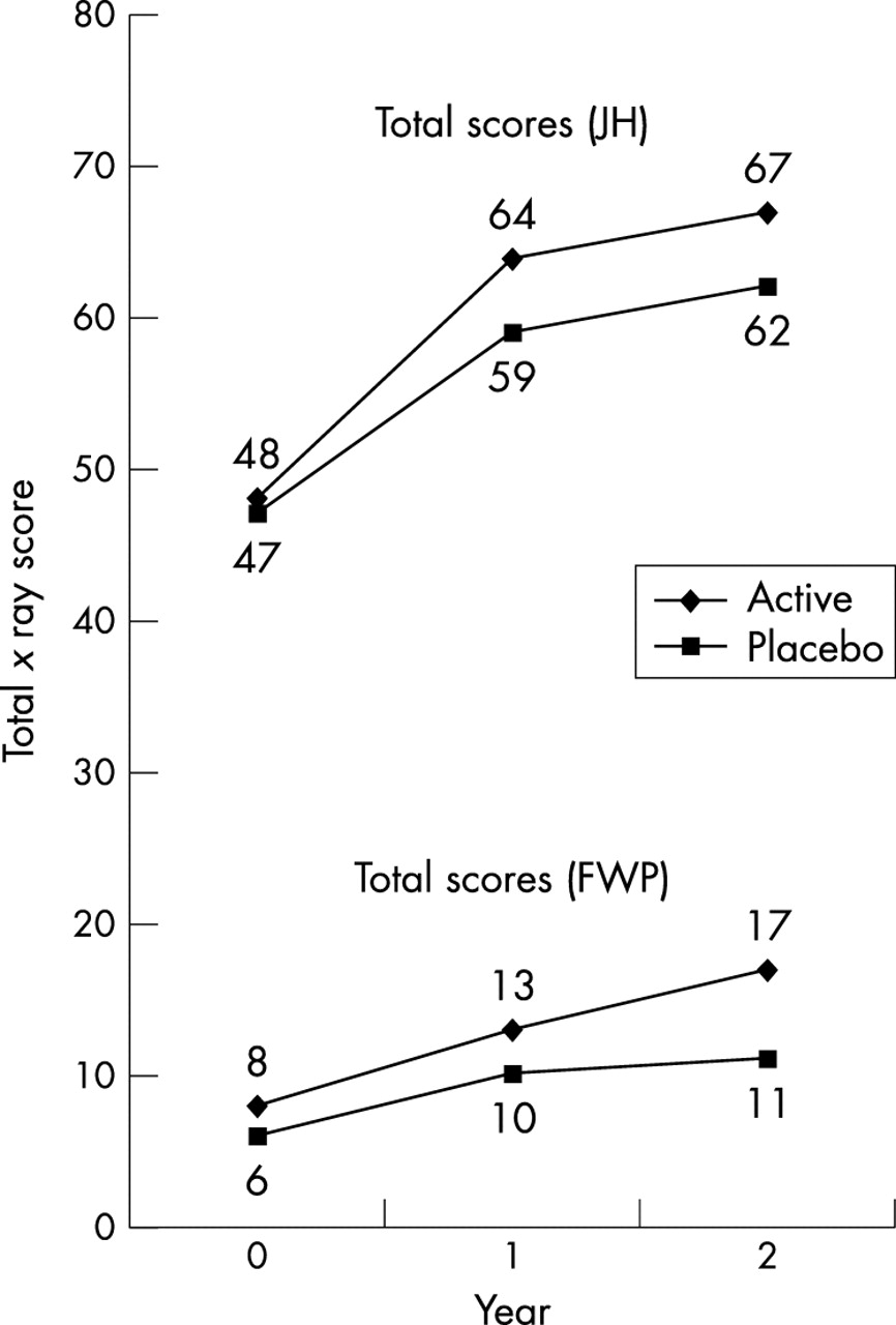
Radiology was available in 64/83 (77%) on active prednisolone and 66/84 (79%) on placebo. The correlation coefficient between the total score for all x rays read in random order and those read in chronological sequence was r = 0.8 (p = 0.01). Twenty three patients had no erosions at the start of the study; eight had developed erosions after two years; two were on prednisolone and six on placebo. Of patients with erosions at onset, 59% and 61% showed progression in the placebo and prednisolone groups, respectively. Comparison of initial and two year total scores in both placebo and prednisolone groups showed significant deterioration (table 2). There was no difference between the groups in total scores or in any other radiological variable examined individually for both random (JH) and chronological (FWP) scoring (table 2, fig 2). The results were similar when reading hands and feet separately and when scoring erosions/narrowing separately (data not shown).
Radiological data (van der Heijde modification of Sharp method)

Changes in total x ray score over two years. FWP, F W Poon (radiologist), chronological scoring; JH, J A Hunter (rheumatologist), random scoring.
Clinical efficacy
There was an improvement in clinical measures and in a “modified ACR” (20% improvement in Ritchie articular index and in four of five variables (ESR, pain score, physician global, patient global, HAQ24) in both groups. At one year, 53% of prednisolone treated patients showed a “modified” 20% response, compared with 39% on placebo (p = 0.07). At two years, 41% on prednisolone and 43% on placebo showed a “modified” ACR 20% response. The group receiving oral prednisolone had a lower ESR at year 1 (p = 0.05) but not at year 2. There were no other significant differences between the groups at years 1 or 2 in other clinical or laboratory measures or in functional ability. The results are detailed in table 3.
Clinical and laboratory change in the two groups
Toxicity
Toxicity data relating to blood pressure, weight, BMI, and DEXA are shown in table 4.
Toxicity data relating to blood pressure, weight, body mass index, and bone mineral density over two years
Antihypertensive and anti-bone-resorptive treatment was more often used in the prednisolone group (table 5). Despite this there was a trend towards higher diastolic blood pressure in the active group at year 1 (table 4). There was also a significant weight gain in the active group in years 1 and 2, with an associated increase in BMI. The placebo group did not show an increase in weight at year 1 but there was a borderline change at year 2. There was no significant difference in the lipids at outset or at 2 years in those patients in whom it was measured (total cholesterol/HDL cholesterol).
Concomitant treatment
The percentage change in femoral neck and L2–L4 DEXA scores in the active prednisolone and placebo groups were compared using the Mann–Whitney U test. There was no significant difference between the two groups for either score (p = 0.315 and 0.215, respectively).
Concomitant treatment
Concomitant treatment is shown in table 5. Use of non-steroidal anti-inflammatory drugs (NSAID) decreased in both groups to a similar extent, in keeping with a DMARD response. Low dose aspirin and treatment for ischaemic heart disease remained similar, whereas the use of antihypertensive agents increased in both groups, as did prescription of lipid lowering agents. The use of any treatment for osteoporosis also increased in both groups.
Additional intramuscular or intra-articular corticosteroids
Additional intramuscular or intra-articular steroids were given to 43% of the placebo group and 49% of the active group over the course of the study (mean dose 55 mg of triamcinolone acetate and 52 mg in placebo and active groups, respectively).
DISCUSSION
The most striking finding in this study was that there was no demonstrable benefit of low dose prednisolone on progression of erosions, a marker of bone damage, or on reducing the extent of joint space narrowing, an indicator of cartilage loss, as noted on x rays of hands and feet. This was true when x rays were read in chronological or totally random order. Low dose prednisolone provided marginal clinical benefit in early rheumatoid arthritis but this was not sustained beyond 12 months. This is similar to the results of other studies and suggests that when used with early DMARD treatment oral prednisolone provides no additional sustained advantage in symptoms or in measures of the inflammatory component of the disease.
Of the previous studies that have shown radiological benefit from low dose oral corticosteroid therapy, the largest and most rigorous is the ARC multicentre study. Scrutiny of factors that might explain the discrepancy in results reveals that conventional DMARD use was more consistent in WOSERACT than in the ARC study. All the patients in WOSERACT received sulphasalazine initially (and a subsequent DMARD in 14% if sulphasalazine was not tolerated), whereas only 66% of patients in the ARC study received a DMARD and there was no consistency across the two treatment allocations in that study. It is possible—although there is no evidence base—that sulphasalazine does not synergise with corticosteroid treatment. The female to male ratio, age distribution, disease duration, clinical disease severity, and rheumatoid factor positivity were similar (acute phase response and rheumatoid factor status28,29 have been shown to have an influence on radiological progression), and the proportion of patients in whom x rays were available was also similar (83% in the ARC study and 78% in ours). The studies differed in that the proportion of patients with erosive disease was different in WOSERACT from the ARC study. Significantly more patients in our study had erosive disease (71% had erosive disease of hands in this study, compared with 31% in the ARC study, p = 0.03 by χ2; the data were similar at the end of year 1). However, in the ARC study more patients in the placebo group compared with the prednisolone group had erosive disease at the outset, whereas the WOSERACT groups were well matched, probably as a result of randomisation with stratification using a minimisation technique.
The natural history of non-erosive rheumatoid disease is well documented—few patients without erosive disease at the time of diagnosis progress. This contrasts with those who had erosive changes at the start of the study; in these the progression was the result of damage increasing in the eroded joints over time, rather than more non-eroded joints developing erosions. It is therefore possible that the difference in results partly reflects the populations studied, with a bias towards erosive disease in the WOSERACT cohort. Long term more severe disability is more likely to develop in such patients.
We used a different method to measure x ray damage in WOSERACT than was used in the ARC study—namely, the van der Heidje modification of the Sharp method rather than the Larsen score.30 JH, using the random method, had higher scores than FWP, who used the chronological method. It was nevertheless reassuring that the change in x ray score was consistent between the two readers. The modified Sharp method is the preferred measurement in evaluating change over time, has a larger range and a greater sensitivity to change, and takes into account changes in the feet, one of the first sites to be affected in rheumatoid arthritis. It has also been the more common method used in recent studies of DMARD in rheumatoid arthritis.31 Furthermore, to be 95% confident to detect true change by the Larsen score an increase in score of 8 units is required over one year; the mean increase in the ARC study was 5.4 units over two years.
An alternative explanation of the divergent results of the two studies may be a genetic difference in the populations studied. We have previously observed that there is an over-representation of HLA-DR4 in the west of Scotland32; this allele is associated with more severe clinical and radiological disease. Differences in corticosteroid metabolism or in the frequency of the CRF gene polymorphisms—which have been shown in one rheumatoid arthritis cohort—may be another explanation that requires further evaluation.
The male to female ratio in this study is unlike many other DMARD studies in that comparatively greater numbers of men than women were willing to accept the risk of steroids. Men comprised 35% of our group, which is considerably more than in the other DMARD studies but similar to that in the ARC and COBRA studies.7,10 It may be that women are more concerned about the perceived cosmetic problems associated with corticosteroids, and were therefore more likely to refuse to take part.
It is noteworthy that the prescription of antihypertensive agents and osteoporosis treatments increased in both groups. Weight gain occurred in the prednisolone group at years 1 and 2. The borderline change in the placebo group might reflect improvement in disease activity. These trends probably in part reflect monitoring of risk factors. While observed toxicity from corticosteroids in terms of hypertension, weight gain, and osteoporosis could be reduced by active assessment and prompt intervention, there is no room for complacency. Such interventions are expensive and long term sequelae cannot all be prevented, as has been highlighted in observational cohorts.
Conclusions
Our study failed to demonstrate any radiological or clinical benefit over two years from low dose prednisolone in patients with rheumatoid arthritis. It is possible that the standardised DMARD (sulphasalazine) and extensive use of alternative DMARDs in patients who failed on sulphasalazine conferred benefits in this study which outweighed any prednisolone effect. These results suggest that the routine use of low dose prednisolone provides no benefit in patients with rheumatoid arthritis receiving DMARD.
APPENDIX
Responsibilities
Study design: Dr H A Capell, Dr R Madhok, Dr J A Hunter, Dr D Porter, and Dr E Morrison.
Study co-ordinator: E A Thomson
Randomisation: A Tierney
X Ray scoring: Dr J A Hunter, Dr F W Poon
Data collection: Rosie Hampson
Data analysis: Dr H A Capell, Dr D Porter, and Dr R Madhok
Manuscript preparation: Dr H A Capell, Rosie Hampson, Dr J Larkin, and Dr R Madhok
Patient recruitment: Dr Maciej Brzeski (Falkirk and District General Hospital); Dr Hilary A Capell, Dr Max Field, Professor Iain McInnes, Dr Rajan Madhok, Professor Roger Sturrock (Glasgow Royal Infirmary); Dr Susan Fraser and Dr Elaine Morrison (Southern General Hospital, Glasgow); Dr Paul McGill (Stobhill Hospital, Glasgow); Dr Elizabeth Murphy (Law Hospital, Lanarkshire); Dr Malcolm Steven (Raigmore Hospital, Inverness); Dr Asad Zoma (Stonehouse Hospital, Lanarkshire).
Acknowledgments
We are grateful to Ann Tierney for typing the manuscript, Dorothy McKnight for computing assistance, and Drs Alistair Forrester and Denis Pitkeathley for reading some x rays. Metrology assistance was provided by Sylvia Armstrong, Fiona McDonald, Darryl McGhee, Fiona McGhee, Elizabeth McIvor, Anne Paterson, and Anne Stirling. Dr A Kelman, University of Glasgow, assisted with the statistical method of minimisation.
The Centre for Rheumatic Diseases receives an integrated clinical arthritis centre grant from the Arthritis Research Council (UK). We are also grateful to The Sir Hugh Fraser Foundation (Glasgow) for financial support to purchase prednisolone and placebo tablets.
REFERENCES
Footnotes
-
↵* West of Scotland Early Rheumatoid Arthritis Corticosteroid Trial