Article Text

Statistics from Altmetric.com
Arrhythmias in pregnancy are common and may cause concern for the wellbeing of both the mother and the fetus. For some mothers the arrhythmias may be a recurrence of a previously diagnosed arrhythmia or the first presentation in a woman with known structural heart disease. In most cases, however, there is no previous history of heart disease, and the new occurrence of a cardiac problem can generate considerable anxiety. The majority of arrhythmias that occur during pregnancy are benign, and simply troublesome; hence, advice about appropriate actions during symptomatic episodes, together with reassurance, is usually all that is required. In the remaining minority of cases, judicious use of antiarrhythmic drugs will lead to a safe and successful outcome for both mother and baby. While there were no documented maternal deaths from primary arrhythmias in the last UK confidential enquiry into maternal mortality,1 9% of cardiac deaths were defined as sudden adult death syndrome, which raises the possibility of death from a primary arrhythmia. In women with known structural heart disease, however, arrhythmia is one of the five independent predictors of having a cardiac event during the pregnancy and should therefore be treated seriously.2
INCIDENCE OF ARRHYTHMIAS IN PREGNANCY
Palpitations are a very common symptom in pregnancy. As cardiac arrhythmias can be identified on Holter recordings in up to 60% of normal people under the age of 40 years, it is not surprising that the antenatal clinic encounters its fair share of palpitations.3 In pregnancy, heart rate (HR) increases by 25%; thus sinus tachycardia, particularly in the third trimester, is not uncommon. Ectopic beats and non-sustained arrhythmia are encountered in more than 50% of pregnant women investigated for palpitations while sustained tachycardias are less common at around 2–3/1000.4–6
There are well known gender differences in the incidence and risk factors of a variety of arrhythmias. Symptomatic atrial re-entrant and atrioventricular nodal re-entry tachycardias have a female predominance, while the opposite is true for atrial fibrillation. Drug induced torsade de pointes and symptomatic long QT syndrome are also more common in females. Pathological bradycardia in pregnant women is rare and usually secondary to congenital heart block, with an incidence of 1:20 000 women.7
MECHANISM OF ARRHYTHMIA
The mechanism of arrhythmia generation is shown in table 1. The cardiovascular system undergoes significant change in adaptation to pregnancy, including an increased heart rate and cardiac output, reduced systemic resistance, increased plasma catecholamine concentrations and adrenergic receptor sensitivity, atrial stretch and increased end-diastolic volumes due to intravascular volume expansion, as well as hormonal and emotional changes. A combination of these and the heightened visceral awareness experienced in pregnancy may lead a woman to seek advice on symptoms that are within the normal range and may otherwise have been ignored. The pregnant state is unlikely to generate a new arrhythmia substrate, however; such physiological changes may render a pre-existing substrate capable of sustaining an arrhythmia. A bigger heart can sustain re-entry more easily and mechanical stretch is well known to be arrhythmogenic. Pregnancy may also affect the trigger for arrhythmia generation. Most tachycardia episodes are initiated by ectopic beats and the occurrence of arrhythmia episodes may therefore increase during pregnancy in line with the increased propensity to ectopic activity.
ARRHYTHMIA DIAGNOSIS
There are three elements to the diagnosis of arrhythmia in pregnancy: firstly, obtain an accurate diagnosis of the arrhythmia in order to give a reliable opinion about the prognosis and appropriate treatment; secondly, determine whether or not there is additional heart disease associated with the arrhythmia; finally, exclude systemic disorders that may present with arrhythmias—for example, abnormalities of thyroid function should always be excluded, while hemorrhage, pulmonary embolism, infections and inflammatory states must be considered in cases of unexplained sinus tachycardia or atrial fibrillation.
A thorough history and examination of the patient is paramount. It should be remembered that in the third trimester, patients may become more symptomatic with activity and thus even minor arrhythmias may present with associated symptoms such as breathlessness or chest pain. Palpitations are the most common symptom and while syncope or pre-syncope may reflect a cardiac cause, it may also result from the physiological drop in blood pressure because of peripheral vasodilatation that is maximal in the second trimester. A thorough family history concentrating particularly on any sudden premature or unexplained deaths that may identify a genetic propensity to life threatening arrhythmias is also important.
Identifying the presence of structural heart disease increases the likelihood of arrhythmias and these are more likely to be significant. Conditions such as mitral stenosis may present for the first time during pregnancy, particularly as pregnancy is often the first time a woman is examined by a healthcare professional since childhood. Women with congenital heart disease, particularly those having undergone surgery, are particularly vulnerable to arrhythmias, which may be haemodynamically compromising and warrant special consideration. Patients who had atrial surgery, such as a Mustard procedure for transposition or a Fontan procedure, are particularly vulnerable to atrial flutter, as are those with any cause of right ventricular impairment. Patients who have undergone surgical correction of tetralogy of Fallot may experience atrial flutter or ventricular tachycardia arising from the right ventricular outflow tract, particularly if the correction was incomplete and there is a residual haemodynamic abnormality.
THE RESTING ECG
The resting ECG of a pregnant woman may be slightly different to that of a non-pregnant woman.8 The increase in heart rate may lead to decreased PR, QRS and QT intervals; however, there is usually no change in the amplitude of the P wave, QRS complex or T wave. The electrical axis may shift to the left because of the gravid uterus, and ectopics (premature atrial/ventricular beats) are common during pregnancy. There may be a Q wave and inverted T wave in the inferior leads. The resting ECG may have abnormalities indicative of primary electrical disease such as delta waves of Wolff–Parkinson–White (WPW), or may reflect an underlying cardiac condition or surgical intervention received in the past.
PROLONGED ECG RECORDING
Ideally, a 12 lead ECG should be recorded during symptoms, although this is rarely achieved. Because of their spatial pattern, paroxysmal arrhythmias are the most difficult to diagnose. They can be short lived and the majority have no pattern, so attempting capture with a routine 24 h Holter monitor is often fruitless. Detecting the arrhythmia predominantly depends upon its frequency. If it occurs on a daily basis then a 24 h or 48 h Holter may be sufficient. It is crucial for the woman to keep an accurate diary so any symptoms can be related to any abnormality on the Holter. Asymptomatic arrhythmias should not be treated unless felt to be life threatening. Episodes which are less frequent or which have evaded detection are best documented using a patient activated event recorder. It is important that the woman continues with her normal activity while wearing the device and keeps a diary of her symptoms, which can be related to the recorded rhythm. Finally, implantable loop recorders are increasingly being used, particularly in patients with unexplained syncope. There is no experience of these devices in pregnancy; however, there are no theoretical contraindications to their use.
Table 2 lists the diagnostic modalities used in the diagnosis of arrhythmia during pregnancy.
Antiarrhythmic drugs
The decision to treat a woman depends upon the frequency, duration and tolerability of the arrhythmia. It is a balance between the benefit of arrhythmia reduction or termination and the maternal and fetal side effects of any drug treatment. The greatest risk to the fetus is during organogenesis and this is complete by the end of the first trimester. The smallest recommended dose should be used initially and be accompanied by regular monitoring of maternal and fetal clinical condition. Various drugs have been used to terminate fetal arrhythmias, providing useful safety data. However, the literature is limited to single or small case series. These include digoxin, adenosine, amiodarone, flecainide, procainamide, propranolol, propafenone, quinidine, sotalol and verapamil. The majority of drugs available, however, only have class C evidence for use in pregnancy. Therapeutic levels of drugs may also be difficult to maintain in the pregnant woman as pregnancy tends to decrease the concentration of drugs because of an increased volume of distribution and increased drug metabolism. This may explain why women previously stable on therapy have breakthrough arrhythmias during pregnancy. Table 3 lists the available antiarrhythmic drugs and their complications during pregnancy.
Historically, there has been concern regarding the effect of β-blockers and intrauterine growth restriction (IUGR). This effect seems to be limited to atenolol and when used either at the time of conception and/or during the first trimester.9 10 It was not seen with other antihypertensives used in the first trimester, or when atenolol was taken in the second or third trimester only. Therefore, where benefit exceeds risk—for example, arrhythmias, mitral stenosis, Marfan syndrome, thyrotoxicosis—β-blockers should not be withheld.
DC cardioversion
DC cardioversion is safe in all stages of pregnancy; because the amount of current reaching the fetus is small, it is only associated with a small risk of inducing fetal arrhythmias. There have been reports of the need for emergency caesarean section because of fetal arrhythmia, particularly in women who are compromised; hence the fetus should be carefully monitored before and throughout the procedure. In the latter stages of pregnancy, some anaesthetists prefer to carry out the procedure using full general anaesthetic and intubation in view of the more difficult airway and increased risk of gastric aspiration. Women should be nursed with the pelvis tilted to the left to relieve caval compression, but otherwise the procedure is the same as for non-pregnant women.
Implantable cardioverter-defibrillators
Women with implantable cardioverter-defibrillators (ICDs) have successfully negotiated pregnancy with good fetal outcomes. One study reported a series of 44 pregnant women with an ICD implant in situ and found no increases in either device or treatment complications, nor any increase in the number of shocks the women received compared to preconception.11 There were no deaths and only one still birth despite eight of the women receiving one or more shocks during their pregnancy. In addition to standard ICD management, after each therapy, monitoring of the baby is advised. The electrical field to which the fetus is exposed is minimal; however, if the arrhythmia was associated with hypotension, placental blood flow may have been compromised leading to fetal distress. For those pregnant women with ongoing malignant arrhythmias despite pharmacological treatment, an ICD may be a safe alternative. The prompt detection and treatment may reduce haemodynamic instability and some groups have reported successful implant using echocardiography to guide the positioning of the leads and avoiding radiation exposure to the fetus.
MANAGEMENT OF SPECIFIC ARRHYTHMIAS
Bradycardia
Pathological bradycardia in pregnant women is rare. Some women who have physiological bradycardia may, in the second trimester, feel symptomatic as their blood pressure drops due to a reduction in systemic resistance; however, treatment is rarely required. Rarely, symptomatic bradycardia has been attributed to supine hypotensive syndrome of pregnancy, which is a result of compression of the inferior vena cava by the gravid uterus and responds to maternal changing of position. Congenital heart block is rare and does not usually pose a problem during the pregnancy. There is conflicting advice about whether to advise temporary pacing for delivery. Proponents state concern that the Valsalva manoeuvre associated with delivery increases the chance of worsening bradycardia and syncope, and pacing also allows for an adequate heart rate response for the increased cardiovascular stress. The alternative view is that, from experience, patients rarely use the device that has been inserted.12 Spinal anaesthesia for caesarean section, however, can be associated with a higher incidence of all grades of bradycardia (up to 13%).13
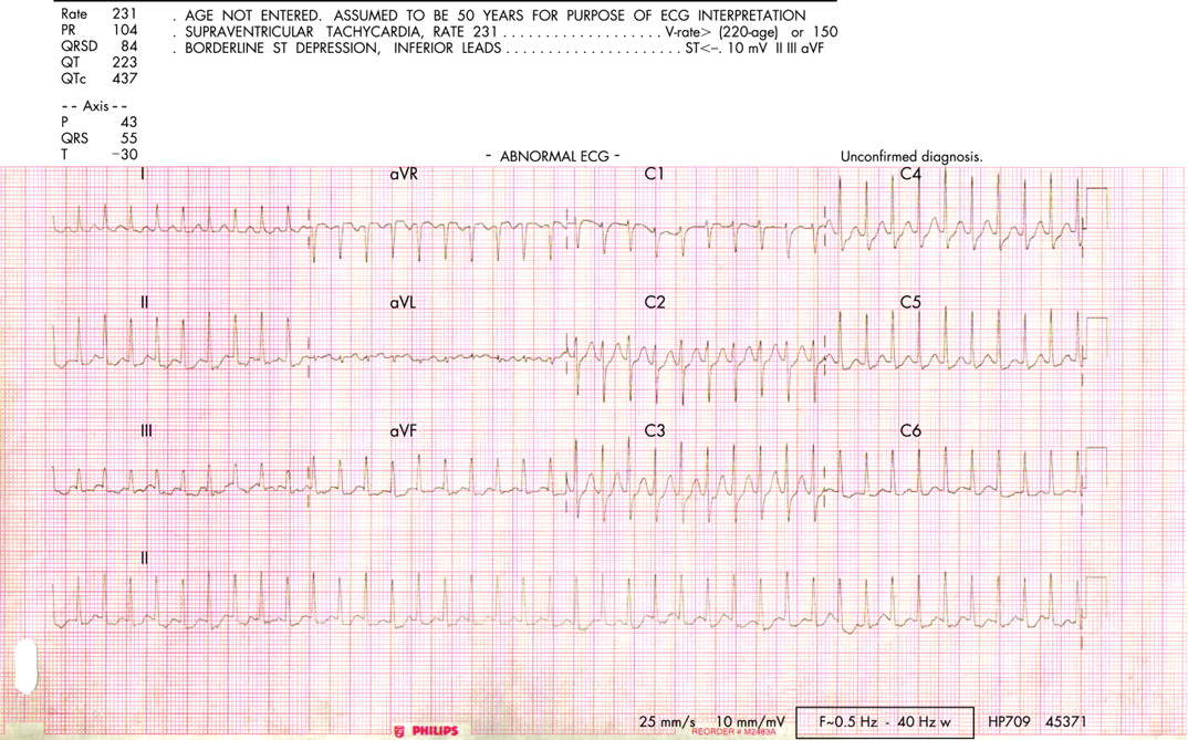
Supraventricular tachycardia
An example of supraventricular tachycardia (SVT) is shown in fig 1. The drug of choice is partly dependent upon the SVT being treated. Analysis of the onset and termination of the arrhythmia, the P wave morphology and relationship of the P wave to the QRS complex, and the response to adenosine, will often allow differentiation between different SVT mechanisms, but this is not always the case. Whatever the precise mechanism, vagotonic manoeuvres such as carotid sinus massage may terminate the episode, and thus should be attempted. If successful they can be easily self administered by the patient to deal with recurrences. When vagotonic manoeuvres fail, intravenous bolus adenosine can be used, with escalating boluses up to a maximum of 18–24 mg until the desired response is achieved. While an increase in intravascular volume occurs in pregnancy, the enzyme responsible for degradation of adenosine, adenosine deaminase, decreases and therefore most women respond to doses between 6–12 mg. As the half life of adenosine is so short, it does not effect the fetus. However, as it may encourage conduction down an accessory pathway and accelerate an arrhythmia, it is recommended that it is administered by experienced personal in a monitored area with equipment available for resuscitation. Verapamil is an effective second line treatment for the treatment of SVTs and can be used in doses up to 10 mg without affecting fetal heart rate, though fetal distress has been associated with verapamil induced maternal hypotension. β-blockers are the drugs of choice in women with known WPW, as atrioventricular (AV) nodal blocking drugs such as digoxin and calcium channel blocking drugs may accelerate conduction through the accessory pathway and cause a deterioration in maternal condition.

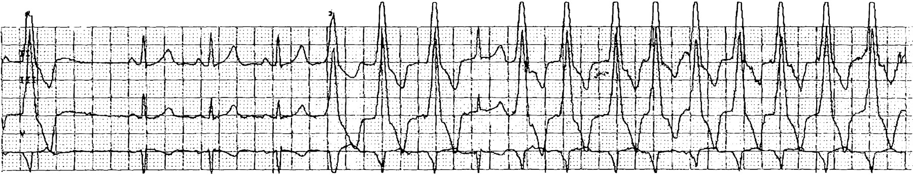
Atrial fibrillation and flutter
Atrial fibrillation and flutter (fig 2) are uncommon in pregnancy; if seen, however, they are most commonly associated with congenital or valvular heart disease as well as metabolic disturbances such as thyrotoxicosis or electrolyte disturbance. Though often well tolerated apart from in severe mitral stenosis, it is advantageous to terminate the arrhythmia to avoid the need for anticoagulation, particularly as pregnancy is a prothrombotic state. Preferred drugs are β-blockers (sotalol, atenolol) or flecainide. Procainamide is another safe alternative, while mexiletine and amiodarone have also been used in small numbers of cases in the acute setting with success. In those where rate control is required rather than restoration of sinus rhythm, β-blockers, verapamil and digoxin are the preferred drugs along with adequate anticoagulation.

If there is impaired ventricular filling such as mitral stenosis or diastolic ventricular dysfunction, the high ventricular rates associated with atrial fibrillation or flutter will reduce ventricular filling because the proportion of the cardiac cycle spent in diastole decreases as the heart rate increases. This results in an increase in filling pressures and reduced forward flows, together with peripheral vasoconstriction and reflex salt and water retention. Unless checked, this process may rapidly progress to acute pulmonary oedema. The intense catecholaminergic drive that occurs in pulmonary oedema further increases the heart rate and filling pressures. As well as the usual treatment for pulmonary oedema of oxygen, diuretics, morphine and nitrates, a β-blocker—given intravenously if necessary—is essential and may be life saving in this situation.14
Idiopathic ventricular tachycardia
Idiopathic right ventricular tachycardia (VT) originating from the right ventricular outflow tract just below the pulmonary valve is the most common form of idiopathic VT seen in pregnancy (fig 3). It virtually never accelerates to an unstable rhythm and patients most commonly experience ectopic beats, bigemini or short runs of non-sustained VT. The QRS morphology is of a left bundle branch block pattern with an inferior axis and responds well to β-blockers. Idiopathic left VT with its right branch block pattern is far less common and responds well to verapamil.

Monomorphic ventricular tachycardia in the structurally abnormal heart
Any disease process that affects the ventricular myocardium causing scarring, hypertrophy or infiltration may disrupt the electrical integrity of the myocardium. Rapid VT causes hypotension, reduced myocardial coronary perfusion and subendocardial ischaemia, an unstable situation that may degenerate into ventricular fibrillation. VT in the presence of structural heart disease is associated with a significant risk of sudden death and requires emergency treatment. Treatment should be with intravenous lignocaine, amiodarone or DC cardioversion.
Polymorphic ventricular tachycardia
Unless polymorphic VT spontaneously terminates within a few seconds, it invariably causes collapse and has a high risk of degenerating into ventricular fibrillation. The emergency treatment requires correction of electrolyte disturbance including magnesium, removal of precipitating drugs, particularly class I and III antiarrhythmic drugs, macrolide antibiotics, non-sedating antihistamines, antidepressants and some antipsychotics, and potentially temporary overdrive pacing.
Cardiac arrest
This is rare (1:30 000 deliveries); however, if it occurs it is important to be aware of a few differences in the cardiopulmonary resuscitation techniques of the pregnant woman.15 In order to optimise fetal outcome, resuscitation should proceed following established guidelines from the resuscitation council and an obstetrician and paediatrician should be involved from an early stage. It is important to remember, in addition to the standard causes of cardiac arrest, amniotic fluid embolism, pulmonary embolism, peripartum cardiomyopathy and acute coronary or aortic dissection are important causes in the pregnant or recently delivered woman. In the later stages of pregnancy, the uterus can reduce venous return because of aorto-caval compression, particularly in the supine position. There are a number of reports where relief of this by emergency caesarean section has been associated with a successful resuscitation of the mother. This obstruction can also be relieved using sandbags or a “Cardiff wedge” under the right side of the patient, manual displacement of the uterus to the left or raising of the mother’s right hip. Chest compression may be more difficult because of the enlarged breasts and the splinting of the diaphragm. Gastric emptying is delayed in pregnancy and so early intubation is recommended to prevent aspiration. Peri-arrest caesarean section is performed to aid resuscitation of the mother rather than to “save” the baby.
CONCLUSION
Pregnancy may be associated with various types of ventricular and supraventricular arrhythmias. Ideally, management should start before conception, but during pregnancy treatment should only be initiated for severe symptoms or haemodynamic compromise.
In general, arrhythmias during pregnancy can be safely managed medically with little risk to mother or fetus. Drugs should be avoided in the first trimester if possible and the choice of drug should reflect its safety record in pregnancy as well as the particular arrhythmia being treated and any associated structural heart disease. Drugs should be administered in the lowest effective dose and the mother and fetus monitored carefully throughout the pregnancy.
In emergency situations, or where medical treatment has failed, DC cardioversion can be safely administered throughout pregnancy. Cardiac arrest is rare; however, the physician should be aware of the special circumstances that need to be considered.
INTERACTIVE MULTIPLE CHOICE QUESTIONS
This Education in Heart article has an accompanying series of six EBAC accredited multiple choice questions (MCQs).
To access the questions, click on BMJ Learning: Take this module on BMJ Learning from the content box at the top right and bottom left of the online article. For more information please go to: http://heart.bmj.com/misc/education.dtl Please note: The MCQs are hosted on BMJ Learning—the best available learning website for medical professionals from the BMJ Group.
If prompted, subscribers must sign into Heart with their journal’s username and password. All users must also complete a one-time registration on BMJ Learning and subsequently log in (with a BMJ Learning username and password) on every visit.
Full explanations for the answers to the MCQs accompanying this article are available from the Heart office—HeartJournal@BMJGroup.com.
Other articles of use
Silversides CK, Harris L, Haberer K, et al. Recurrence rates of arrhythmias during pregnancy in women with previous tachyarrhythmia and impact on fetal and neonatal outcomes. Am J Cardiol 2006;97:1206–12.
Nelson-Piercy C. Handbook of obstetric medicine, 3rd ed. London: Taylor Francis (Informa Health Care), 2006. A pragmatic guide to the management of medical problems in pregnancy.
Adamson DL, Dhanjal MD, Nelson-Piercy C, et al. Cardiac disease in pregnancy. In: Greer, Nelson-Piercy, Walters, eds. Maternal medicine. Elsevier, 2007:14–39. A chapter reviewing management of all aspects of cardiac disease in pregnancy including anaesthetic considerations.
REFERENCES
Footnotes
Both authors also at Department of Obstetric Medicine, Queen Charlottes and Chelsea Hospital, London, UK
In compliance with EBAC/EACCME guidelines, all authors participating in Education in Heart have disclosed potential conflicts of interest that might cause a bias in the article.