
Background
Human embryonic stem cells (hESCs) are pluripotent cells that originate from the inner cell mass of a blastocyst. Their limitless differentiation capacity and ability to self-renew, also known as ‘stemness’, give them great potential in regenerative medicine. However, hESCs need to be maintained as a highly proliferative and pluripotent population to be used in clinical application.1 Much evidence suggests that a hypoxic (5% oxygen) culture is more beneficial in maintaining hESC stemness than atmospheric (20% oxygen) cultures.1–4 Studies have shown a higher expression of transcription factors NANOG, SOX2 and OCT-4, which regulate hESC pluripotency, and a lower rate of apoptosis in hESCs cultured at 5% oxygen compared with those maintained at 20% oxygen. This is perhaps not surprising since the pre-implantation embryo develops in a hypoxic reproductive tract, ranging from 1.5–5.3% oxygen concentration.5
However, the regulation of apoptosis in hESCs under hypoxia has been largely overlooked.6 BCL2 anthogene 1 (BAG1) is a recently discovered multifunctional protein that is involved in many cellular processes including apoptosis, transcription, proliferation, cell signalling and differentiation.7 BAG1 expression has been shown to be upregulated in mesenchymal and trophoblastic cells under hypoxia, suggesting that its expression may be regulated by environmental oxygen tensions.7 BAG1 is overexpressed in many cancers, suggesting that it may contribute to cancer pathogenesis by increasing resistance to apoptosis.8 BAG1 knockout experiments in mice have demonstrated its requirement for the survival and differentiation of haematopoietic and neuronal cells.8
This study therefore aimed to investigate whether BAG1 has a role in regulating the increased proliferation of hESCs observed at hypoxic conditions and whether it has an effect on hESC self-renewal.
Methods and results
Immunocytochemistry showed that BAG1 is expressed in hESCs, suggesting that it may have a function in the maintenance of hESC stemness. It showed that BAG1 expression was localised to both the cytoplasm and nucleus in hESCs cultured at both 5% and 20% oxygen. But Western blotting showed no significant difference in BAG1 expression between hESCs cultured at 5% oxygen tension and those maintained at 20% oxygen tension (Fig 1). This suggests that BAG1 expression in hESCs is not dependent on environmental oxygen tensions.
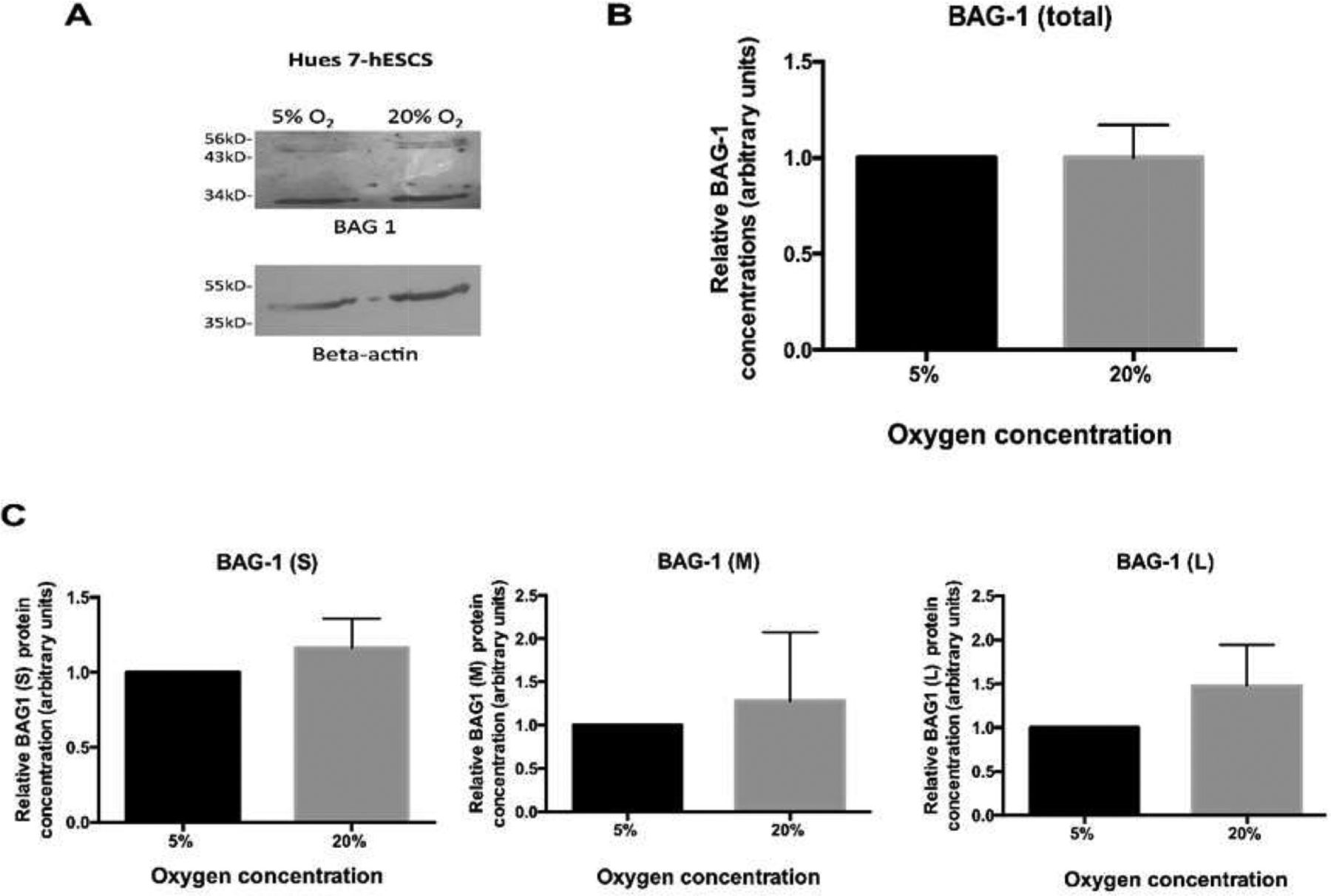
BAG1 expression is not significantly different in hESCs cultured at hypoxia compared with hESCs maintained at atmospheric oxygen. A) Representative blot where data were normalised to β-actin and to 1 for expression at 5% O2. B) No significant difference in total BAG1 expression. C) No significant difference in BAG1 S, M and L expression (n=5). Bars represent mean ± standard error of the mean.
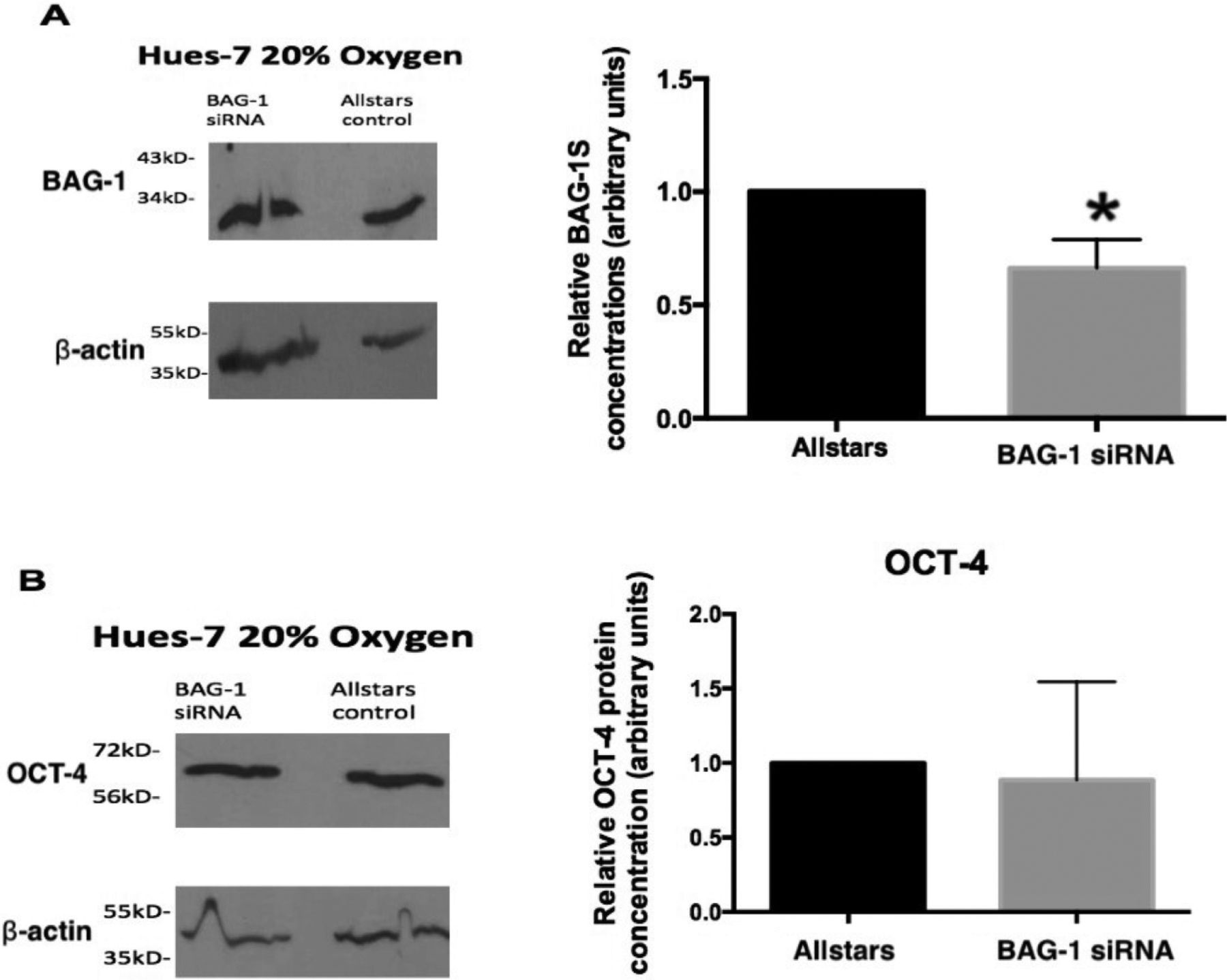
BAG1 was also silenced in hESCs cultured at 20% oxygen using small interfering ribonucleic acid, which showed no significant effect on OCT-4 expression in hESCs compared with negative control (Fig 2). This suggests that BAG1 does not regulate hESC self-renewal. A recent publication by Tang et al (2017) supported this finding in murine embryonic stem cells (mESCs), and reported that BAG1 homozygous knockout mESCs had a normal karyotype and maintenance of pluripotency.9

BAG1 does not regulate OCT-4 expression. Human embryonic stem cells cultured at 20% O2 were transfected with either an Allstars control small interfering ribonucleic acid (siRNA) or BAG1 siRNA. A) BAG1 expression in human embryonic stem cells transfected with BAG1 siRNA was significantly reduced (p=0.0435; n=3). B) No significant difference in OCT-4 expression in BAG1 siRNA-transfected human embryonic stem cells (n=3). Data were normalised to β-actin and to 1 for control siRNA-treated cells. Bars represent mean ± standard error of the mean.
Conclusions
The present study therefore shows that BAG1 does not play a role in the hypoxic response of hESCs and that perhaps other pro-survival proteins may be involved. Gene expression profiling comparing anti-apoptotic gene expression, including BAG1, between hESCs and cancer cell lines showed that only two anti-apoptotic factors, BCL10 and BIRC5 (survivin), are preferentially expressed in hESCs.10 Future work should therefore be directed to elucidating the roles of other anti-apoptotic proteins in hESCs.
Conflicts of interest
None declared.
- © Royal College of Physicians 2020. All rights reserved.


Jump to section
Related Articles
Cited By...
- No citing articles found.